Article • Evolving technique
Flow cytometry rises to new challenges
Flow cytometry has proved an invaluable diagnostic tool for leukaemia and lymphoma for almost three decades. Now, however, this is evolving in applications to seek out residual disease in cases and in fusion with molecular testing to advance its diagnostic potential.
Report: Mark Nicholls
However, although recognised as fast, flexible and accurate, flow cytometry suffers from a lack of standardisation, according to Professor Brent L. Wood from the University of Washington (UW) School of Medicine in Seattle. Wood addressed those issues when describing the impact of flow cytometry on the clinical laboratory at the joint European Federation of Clinical Chemistry and Laboratory Medicine/European Union of Medical Specialists (EFLM/UEMS) conference, which was held this October in Turkey.

Flow cytometry is a technology that analyses the physical and chemical characteristics of particles in a fluid as it passes through a laser. Wood, who directs UW Medicine’s Haematopathology and SCCA Pathology, could reflect on his experiences with flow cytometry for over 20 years. He has been involved in developing flow cytometry in various contexts, such as identifying residual leukaemia after therapy and demonstrating its clinical utility and significance in assessing patient outcome. Flow cytometry has been a technique for diagnosis since the 1970s, particularly of neoplasms, and came to the fore in clinical laboratories in the 1980s with the spread of HIV before evolving as a methodology in the 1990s for diagnostics of leukaemia and lymphoma and diseases that were easier to create cell suspensions from such as peripheral blood and bone marrow. Recently, this advanced to focus on next generation sequencing and examining the concordance and relative merits of those two techniques.
Interpreting data is the subjective component of this type of analysis, which requires someone with experience and training
Brent L Wood
As a technology, flow cytometry is flexible, facilitating the design of a variety of different assays to create panels of re-agents that are suitable for diagnosing leukaemia and lymphoma. ‘The application to monitoring for residual disease involves looking for much smaller populations of cells of similar type and distinguishing them from their normal counterparts,’ Wood said. ‘That’s one of the thing’s that this technology is particularly adept at doing – distinguishing normal from abnormal.’
However, he emphasised that looking for relatively small populations of cells is a more technically-demanding process, requiring more attention to detail in terms of technique, consistency and artefacts, and interpretation. An area of concern, said Wood, is that while the technology is widespread, it is ‘very poorly standardised’. This can see different laboratories having their own panel of reagents that they have validated themselves and their own ways of processing samples and collecting and interpreting data. ‘Interpreting data is the subjective component of this type of analysis, which requires someone with experience and training,’ he pointed out. ‘In that sense it is different to many other tests we do in clinical laboratories, where instruments provide a numerical data or results that are reviewed and released.’

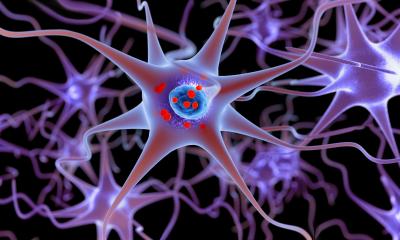
Source: shutterstock/paulista
Steps are being taken to address standardisation; for example, the Euroflow group is working to create standardised panels of reagents in Europe and has made some progress. The issue, he said, is more problematic in the USA, with little incentive for labs to adopt such panels, although an exception has been in minimal residual disease testing, where the FDA has required standardised testing for certain clinical trials. ‘Some manufacturers are beginning to make reagent combinations that are premixed in a stabilised format, so that makes it easier for people to bring that data and to standardise them,’ Wood said. ‘That may have been a step forward,’
Where flow cytometry shows a clear advantage is its speed. ‘When a sample arrives in the laboratory,’ Wood explained, ‘it can be processed rapidly and results are generated in a few hours; that’s difficult to match with antibody-based techniques that require a more lengthy process. It is also inherently multi-parametric with its ability to look at large numbers of individual single cells very rapidly in a multi-parametric fashion, and so provides a high degree of sensitivity and specificity for the identification of abnormal hematic cell populations.’ As for the future, Wood believes flow cytometry technology is capable of providing more information about the inherent biology of leukaemia and lymphomas. ‘We can begin to understand the end-line biology and which has the markers or antigens associated with a particular response to a therapy or outcomes,’ he explained. ‘There is a potential role to assess the biomarkers as they are identified in a relatively efficient or cost-effective manner, but it requires some discovery of what those markers are to drive the application.’
One of the real opportunities lies in the fusion of flow cytometric technology with molecular testing
Brent L Wood
Another area of potential is in residual disease monitoring, where the assessment of both prognosis or eventual outcome and response to therapy is becoming an application increasingly carried out by a variety of laboratories.’ Of further interest is to extend the technology to solid tumours, although this has been more challenging because preparing samples for them is difficult and there is a limited amount of data known about the expression patterns of proteins and antigens in solid tumours.
‘One of the real opportunities lies in the fusion of flow cytometric technology with molecular testing,’ he said, ‘increasing capability to do single cell molecular-based techniques that can incorporate some of the flow cytometric features to a system identifying cell populations, but also then extensively characterising them at a molecular level and providing much more insight into the biologic nature of subpopulations of cells in any given tumour and potentially their relative response to therapy. ‘I think hybridisation of flow cytometry with molecular testing is an important area going forward,’ Wood concludes.
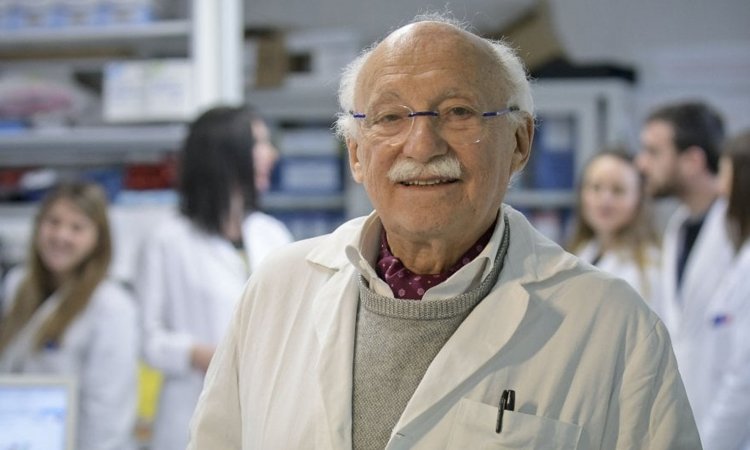
Profile:
Pathologist Professor Brent L Wood is director of UW Medicine’s Haematopathology and SCCA (Seattle Cancer Care Alliance) Pathology laboratories and UW professor of Laboratory Medicine and Pathology. A past president of the International Clinical Cytometry Society, his laboratory serves as one of two national reference laboratories in Canada for flow cytometric identification of minimal residual disease in childhood.
28.11.2018