PET/CT
Morphology and function in the management of heart disease
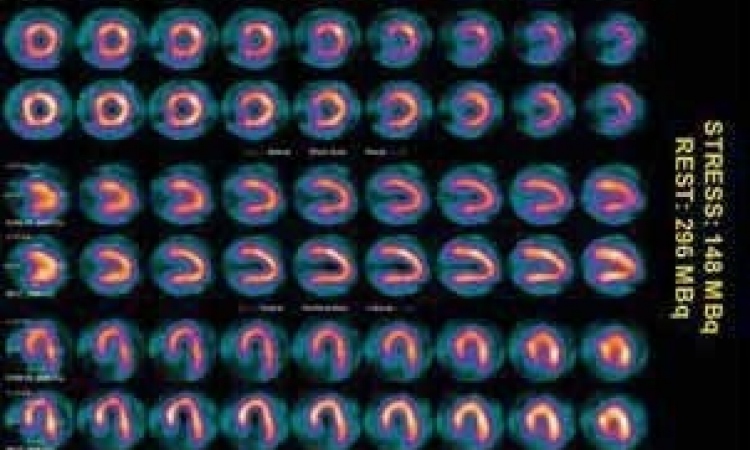
PET/CT combines PET's ability to precisely measure regional myocardial blood flow with the capability of multislice CT imaging.
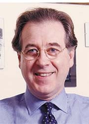
Possibilities and opportunities offered by new PET/CT systems for cardiac research and diagnoses will be an important topic at the ESC in Barcelona this year. During the event, Paolo Camici, Professor of Cardiovascular Pathophysiology at Imperial College School of Medicine, in London, will lecture on this development, which he outlined during an interview with Daniela Zimmermann of European Hospital
The fusion of PET/CT is important because it combines PET’s ability to precisely measure regional myocardial blood flow with the capability of multislice CT to image, non-invasively, the anatomy of coronary arteries, Professor Camici explained: ‘In this way, one can ascertain if a given restriction on the coronary angiogram corresponds to an abnormality of myocardial blood flow in the territory subtended by the diseased vessel. PET is unique,’ he pointed out, ‘because it provides quantitative measurements of myocardial blood flow, that is, in units of millilitres of blood per minute per gram of myocardium. This is at variance with methods that provide only relative measurements based on gradients in tracer uptake between different areas of the heart. With the latter techniques one can only conclude that a given area of the heart has more blood flow relative to another area. Although this approach is clinically useful for the diagnosis of myocardial ischaemia, it is ineffective when blood flow in the whole heart is abnormal. For instance, in patients with cardiomyopathies there are abnormalities of myocardial blood flow that homogeneously affect the whole left ventricle. In these patients, only PET can demonstrate the abnormalities of absolute myocardial blood flow compared with normal volunteers.
‘Furthermore, PET allows us to measure the utilization of important metabolic substrates, such as any free fatty acid by the myocardium. Clinically, these metabolic measurements are used to assess myocardial viability, i.e. the presence of metabolically active tissue within a dysfunctional left ventricular segment.’
What is the potential role of PET/CT in imaging plaques in coronary arteries, for example, to define a type of plaque?
‘At the moment this is a little bit of wishful thinking. There are a lot of technical problems to overcome before we can realise such diagnostic methods. The problem we face is the fact that the heart moves intrinsically as well as secondary to respiratory activity. This motion affects the accuracy of PET/CT measurements and limits the achievable spatial resolution. Unless we find a way to correct PET/CT acquisitions for these movements, the resolution necessary to image plaque cannot be reached. Just imagine: The diameter of a main coronary artery is between 2 and 5 mm, so we are talking about a very small, moving object. To obtain good images you need tracers with a very high specific activity and very high radioactive concentration. We still don’t have any tracers like that. To summarise: specific imaging of coronary plaques by PET/CT is not an application I see in the near future, although a lot of research is happening in this field.’
Does this mean PET/CT could benefit research, but not daily clinical practice?
‘I would not say so. Certainly this combination is very useful for research, but it is also of interest for clinical practice. Clinically, PET has applications for the assessment of viability in combination with FDG and for the diagnosis of coronary artery diseases when used in combination with rubidium-82. Furthermore, PET/CT now allows us to image coronary arteries in a way similar to angiography, but without the need of a catheter.
In your ESC lecture, will highlight any other aspect?
Yes, the fusion of PET and MRI. Taking research aspects into consideration, this will be a very promising combination. MRI can provide a lot of functional parameters without radioactivity, with important ethical advantages. In addition, MRI is very flexible and has a lot of important applications in the fields of cardiology. PET and MRI are very complementary; together they can provide information on regional ventricular anatomy and function, on coronary anatomy and tissue perfusion, on tissue viability and metabolism.
30.08.2006