

Image source: Joy G, Punjabi K, Searle G et al., Lancet Digital Health 2026 (CC BY 4.0)
News • Ejection fraction analysis
AI predicts patient survival from cardiac MRI scans better than clinicians
Findings support use of AI as first-line tool for analysing heart scans for more digitally focused NHS
Artificial intelligence (AI) measurements of heart function from MRI scans are more strongly linked to how long patients live after their scan than measurements made by clinicians, suggesting that AI could provide a useful first-line approach to measuring heart function. This is according to research led by City St George’s, University of London and University College London, and published in The Lancet Digital Health.
The research focused on ejection fraction – the most important measure of heart function. It describes how effectively the heart pumps blood and acts as a ‘gatekeeper’ for many treatments.
Our findings suggest AI can measure [ejection fraction] with remarkable consistency and precision, resulting in a small but meaningful improvement in identifying patients at higher risk
George Joy
Ejection fraction is widely used by doctors to guide treatment decisions for people with heart disease. It helps doctors decide whether patients may benefit from specialist heart failure treatments, implanted heart devices and procedures to improve blood flow to the heart. It is used to gauge surgical risk and to monitor heart function during chemotherapy.
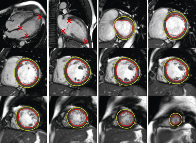
In the largest study of its kind, researchers analysed heart MRI scans from 27,382 patients undergoing investigation for known or suspected heart conditions in the UK and United States. Patients were followed for an average of more than five years, during which 3,458 deaths occurred. The team compared measurements of ejection fraction generated by an AI tool with those produced by experienced clinicians assessing cardiac MRI scans.
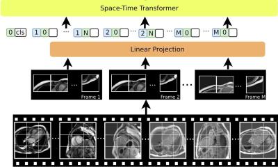
They found that AI-derived measurements of ejection fraction were slightly but significantly better at predicting which patients were at increased risk of death than clinician-derived measurements. The findings remained significant after accounting for factors such as age and sex. The AI tool was also substantially faster, analysing scans in approximately 2-3 seconds per patient. This compared to previous work by the team that found expert clinician analysis typically takes around 13 minutes.
Dr George Joy, NIHR Clinical Lecturer from the Cardiovascular Clinical Academic Group at City St George’s, University of London and lead author, said: "Ejection fraction is one of the most important measurements we make in cardiology because it helps determine how patients are treated and how closely they should be monitored. Our findings suggest AI can measure this important marker with remarkable consistency and precision, resulting in a small but meaningful improvement in identifying patients at higher risk."
The researchers stress that human oversight remains essential. While AI errors were uncommon, with an error rate of around 1% at the patient level affecting just 0.08% of images, they say clinicians should still play a critical role in verifying results and interpreting scans within the broader clinical context.
Dr Rhodri Davies, Associate Professor from University College London (UCL), senior author and inventor of the AI tool, said: "Even modest improvements in accuracy can have a significant impact when applied across thousands of patients. AI is not replacing clinicians, but it can provide faster, more consistent measurements at low-cost and support more streamlined clinical decision-making that ultimately benefits patients.”
The findings support the use of AI as a first-line tool for analysing cardiac MRI scans, with expert review where needed. As healthcare systems face increasing demand, the technology could help reduce workloads while maintaining high standards of patient care and helping patients receive lifesaving treatments sooner.
The team now plan to combine AI measurements from heart scans with those from other tests - particularly electrocardiograms (ECGs) - to spot problems in the heart muscle earlier, so treatment can be initiated before lasting damage occurs.
The research was funded by an NIHR Clinical Lectureship, the NIHR UCLH Biomedical Research Centre and a British Heart Foundation Accelerator Award.
Source: City St George’s, University of London
03.11.2026