© ihorvsn – stock.adobe.com
News • Foreign body aspiration
AI spots hidden objects lodged in patients' airways on CT
Researchers at the University of Southampton have developed an artificial intelligence (AI) tool that can spot hard-to-see objects lodged in patients’ airways better than expert radiologists.
In a study published in npj Digital Medicine, the AI model outperformed radiologists in checking CT scans for objects that don’t show up well on scans. These accidentally inhaled objects can cause coughing, choking, difficulty breathing and sometimes lead to more serious complications if not treated properly. The findings highlight how AI can support doctors in diagnosing complex and potentially life-threatening conditions.
The results demonstrate the real-world potential of AI in medicine, particularly for conditions that are difficult to diagnose through standard imaging
Yihua Wang
The research has been led by Dr Yihua Wang, Dr Zehor Belkhatir, and Prof Rob Ewing at the University of Southampton in partnership with researchers from Wuhan, China. “These objects can be extremely subtle and easy to miss, even for experienced clinicians,” said PhD Researcher Zhe Chen, co-first author of the study from the University of Southampton. “Our AI model acts like a second set of eyes, helping radiologists detect these hidden cases earlier and more reliably.”
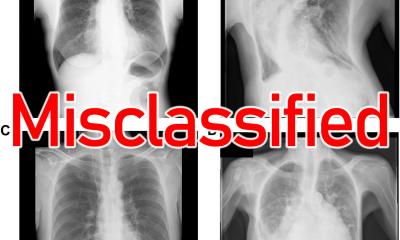
Foreign body aspiration (FBA) occurs when an object, often food or a small piece of material, becomes lodged in the airways. When the objects, such as plant material or crayfish shells, are radiolucent (invisible on X-rays and faint even on CT scans), it can be very difficult to detect. This often leads to missed or delayed diagnoses, putting patients at risk of serious complications. Up to 75% of FBA cases in adults involve radiolucent foreign bodies.
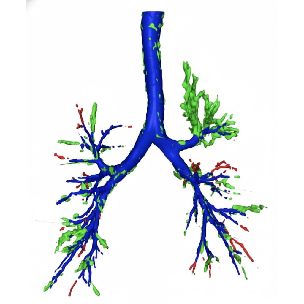
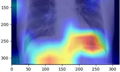
Image source: University of Southampton
To address this challenge, the research team created a deep learning model. It combines a high-precision airway mapping technique (MedpSeg) with a neural network that analyses CT images for hidden signs of foreign bodies. The model was trained and tested using three independent patient groups, consisting of over 400 patients, in collaboration with hospitals in China.
To put the model to the test, researchers compared its performance to that of three expert radiologists, each with over ten years of clinical experience. The task was to examine 70 CT scans, 14 of which were cases of radiolucent FBA, confirmed by bronchoscopy.
When the radiologists detected a case of radiolucent FBA, they did so with total precision – there were no false positives. In comparison, the AI model did so with 77% precision, detecting some false positives. However, the radiologists missed a large portion of FBA cases, identifying just 36% of them and highlighting the difficulty humans have in spotting such cases. The AI model, on the other hand, was able to spot 71% of cases, meaning far fewer FBA cases slipped through the net.
In F1 score, which balances precision and recall, the model outperformed the radiologists with a score of 74% vs 53%. “The results demonstrate the real-world potential of AI in medicine, particularly for conditions that are difficult to diagnose through standard imaging,” commented Dr Yihua Wang, lead author of the study.
The researchers emphasise that the system is designed to assist, not replace, radiologists - providing an additional layer of confidence in complex or uncertain cases. The researchers now aim to conduct multi-centre studies with larger and more diverse populations to improve the model and reduce the risk of bias.
Source: University of Southampton
13.11.2025