Image credit: Osaka Metropolitan University
News • Critical check for deep learning models
AI labeling in radiology: filling in the missing step
Two important checks that make deep-learning models for AI more effective
Researchers at Osaka Metropolitan University have discovered a practical way to detect and fix common labeling errors in large radiographic collections. By automatically verifying body-part, projection, and rotation tags, their research improves deep-learning models used for routine clinical tasks and research projects.
The study was published in European Radiology.
Deep-learning models using chest radiography have made remarkable progress in recent years, evolving to accomplish tasks that are challenging for humans such as estimating cardiac and respiratory function. However, AIs are only as good as the images input into them. Although X-ray images taken at hospitals are labeled with information, such as the imaging site and method, before being fed into the deep-learning model, this is mostly done manually, meaning errors, missing data, and inconsistencies occur, especially at busy hospitals.
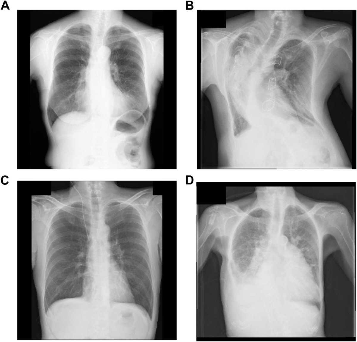
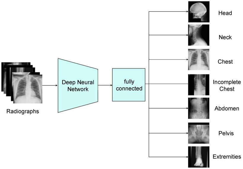
Image source: Mitsuyama Y, Takita H, Walston SL et al., European Radiology 2025 (CC BY 4.0)
This is further complicated by images with various rotations. A radiograph be taken from the anterior to the posterior or vice versa, and it can also be lateral, inverted or rotated, further complicating the dataset. In large imaging archives, these minor errors quickly add up to hundreds or thousands of mislabeled results.
A research team at Osaka Metropolitan University Graduate School of Medicine, including graduate student Yasuhito Mitsuyama and Professor Daiju Ueda, aimed to improve the detection of mislabeled data by automatically identifying errors before they affect the input data for deep-learning models. The group developed two models: Xp-Bodypart-Checker, which classifies radiographs depending on the body part; and CXp-Projection-Rotation-Checker, which detects the projection and rotation of chest radiographs.
Xp‑Bodypart‑Checker achieved an accuracy of 98.5% and CXp‑Projection‑Rotation‑Checker obtained accuracies of 98.5% for projection and 99.3% for rotation. The researchers are optimistic that integrating both into a single model would deliver game-changing performance in clinical settings.
Although the results were outstanding, the team hopes to fine-tune the method further for clinical use. “We plan to retrain the model on radiographs that were flagged despite being correctly labeled, as well as those that were not flagged but were in fact mislabeled, to achieve even greater accuracy,” Mitsuyama said.
Source: Osaka Metropolitan University
06.01.2026