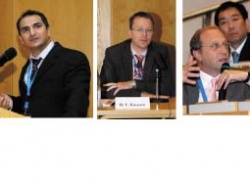
Visualising the breathing lung
After previous meetings in the USA (2002) and Japan (2004), this October the 3rd International Workshop of Pulmonary Functional Imaging (IWPFI) took place in the German Cancer Research Centre, based in Heidelberg University, Germany. " 'The clinicians' need for earlier and more detailed diagnosis in pulmonary disease demands a joint interdisciplinary effort to push the limits in pulmonary functional imaging even further," Professor H U Kauczor, head of the research centre's radiology department and President of the Organising Committee, told participants.

“Over 250 scientists attemded a number far exceeding expectation, which turned the original workshop into a real scientific meeting. As most of the research in this field happens at the interface of medicine and technology, participants came from diverse scientific backgrounds, ranging from Pulmology and Radiology to Medical Physics and Digital Image Processing
and on to Computational Fluid Dynamics. They faced a packed programme of scientific sessions and poster sessions, and two lunchtime symposia sponsored by Toshiba and Siemens.
With this interdisciplinary audience, emphasis was placed on establishing a common basis and terminology for discussion, through keynote lectures from internationally renowned experts presenting important clinical topics and current research activities from different aspects.
Airway disease and emphysema
In his keynote lecture, H Magnussen (Hamburg, Germany) focused on pulmonary function tests, the reference standard, with which functional imaging must compete. On imaging, P Grenier (Paris, France) said that thin-section CT has already proved a reliable method for diagnosis and follow-up of airway disease, thereby already eliminating the need for bronchography. F M Müller (Heidelberg, Germany) consented that CT and MRI already have a place in lung function tests for assessment of early morphological and functional changes in cystic fibrosis. For emphysema, M Mishima (Kyoto, Japan) emphasised the need for new diagnostic methods to describe the different phenotypes of COPD on the way to tailored therapies in the near future.
For quantification, image processing and navigated bronchoscopy, various software tools and programmes were also shown at the industrial exhibition by larger companies alongside research groups presenting competing near-market solutions that incorporate the latest technological advances.
On this, E Hoffman (Iowa, USA) demonstrated how imaging contributes to understand the pathogenesis of emphysema as well as presenting new insights on the interplay of inflammation and hypoxic pulmonary vasoconstriction in the development of emphysema. Moving from perfusion to ventilation, E van Beek (Iowa, USA) presented data on ventilation MRI using hyperpolarised 3He gas, showing good correlation to pulmonary function tests, opening the perspective for supplementary regional information in the near future.
Another method capable of providing information on regional ventilation is oxygen-enhanced MRI, as was reviewed by Y Ohno (Kobe, Japan).
Pulmonary hypertension, exercise testing and biomechanics were another large focus. Multidisciplinary experts discussed the emerging role of imaging – particularly MRI. The challenging application of MRI for such examinations is driven by its unique qualities, no radiation, assessment of function, comprehensive evaluation, potential for repeated, dynamic and stress imaging. For pulmonary hypertension, S Ley envisioned MRI as the single comprehensive imaging modality providing all necessary diagnostic information as a one stop procedure, which he sees at the threshold, because MDCT and MR already provide excellent information on morphology while functional MRI inches up to the so far gold standard echocardiography in functional assessment. Acute pulmonary embolism is a clinical emergency with acute pulmonary hypertension, where modern imaging regimes can not only establish a diagnosis in cases of inconclusive clinical evaluation but also provide prognostic measures, as with obstruction scores and quantification of impaired cardiac function - as summarised in the lectures by groups from Florence, Italy; Lille, France; Toronto, Canada, and Munich, Germany.
Keynotes on ‘Protective Ventilation and Acute Respiratory Distress Syndrome’ included dynamic visualisation of ventilation, recruitment and derecruitment - an exciting concept to tailor ventilation regimens by means of quantitative imaging. In addition, segmented geometries from individual imaging data can now be used to simulate inspiratory and expiratory flow pattern, as well as biomechanical ‘stress’ on the epithelial surface layer.
For oncology, CT now plays a major role for image-guided bronchoscopy, with navigation important to accurately biopsy small nodules, and precisely position brachytherapy probes for endobronchial radiotherapy. To evaluate angiogenesis in lung cancer, imaging was mainly viewed as a possibility to monitor response to therapy with quantitative analysis of tumour perfusion, and to differentiate suspect structures according to their diffusion patterns.
The workshop – The overwhelmingly positive sentiments of participants – and particularly the successful exchange of discussion on highly diverse topics - is a tribute to the organisers wide-ranging vision.”
17.11.2006


