Neurology
Reliving nightmare flight offer new clues about trauma memory
A group of passengers who thought they were going to die when their plane ran out of fuel over the Atlantic Ocean in August, 2001 have had their brains scanned while recalling the terrifying moments to help science better understand trauma memories and how they are processed in the brain.
The neuroimaging study – believed to be an unprecedented examination of a group of people who all experienced the same single blow trauma – is published online today in the journal Clinical Psychological Science (CPS).
Led by Baycrest Health Sciences’ Rotman Research Institute, it is the second major paper to come out in less than a year on passengers who were aboard Air Transat Flight 236 when the crippled plane made a harrowing landing on a small island military base in the Azores to avoid ditching in the ocean.
In the first phase of the study published in 2014 in CPS, the researchers asked the passengers to complete a memory test (without brain scanning) to probe the quality of passengers’ memories of the flight experience three years after the traumatic incident, along with memories of 9/11 and a neutral event. The researchers showed that all the passengers remembered a remarkable amount of detail for the Air Transat incident, regardless of whether or not they had PTSD, although individuals with PTSD tended to veer off topic and recall additional information that was not central to the events assessed.
Almost a decade later, eight passengers agreed to the second phase of the study, which involved brain scanning during presentation of video recreation of the AT incident (obtained from broadcasters such as NBC), footage of the 9/11 attacks, and a neutral event. Of the eight passengers tested, some had a diagnosis of PTSD, but most did not. The group of eight ranged in age from 30s to 60s, including one married couple.
“This traumatic incident still haunts passengers regardless of whether they have PTSD or not. They remember the event as though it happened yesterday, when in fact it happened almost a decade ago (at the time of the brain scanning). Other more mundane experiences tend to fade with the passage of time, but trauma leaves a lasting memory trace,” said Dr. Daniela Palombo, the study’s lead author and currently a post-doctoral researcher at VA Boston Healthcare System and Boston University School of Medicine. “We’ve uncovered some hints into the brain mechanisms through which this may occur.”
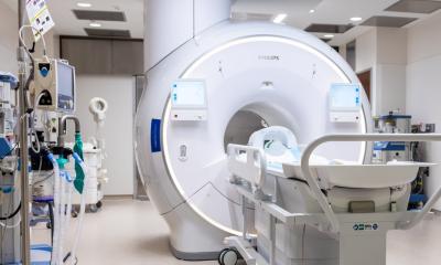
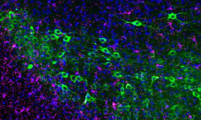
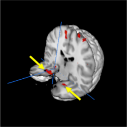
Trauma brain imagePlaced inside a functional magnetic resonance imaging (fMRI) scanner, each of the eight passengers recalled details of their experience on AT Flight 236 while presented with the video clips. Their recollection was associated with heightened responses in a network of brain regions known to be involved in emotional memory – including the amygdala, hippocampus, and midline frontal and posterior regions, when compared to remembering a neutral autobiographical memory.
“Research on highly traumatic memory relies on animal studies, where brain responses to fear can be experimentally manipulated and observed,” said Dr. Brian Levine, senior scientist at Baycrest’s Rotman Research Institute, Professor of Psychology at the University of Toronto, and senior author on the paper. “Thanks to the passengers who volunteered, we were able to examine the human brain’s response to traumatic memory at a degree of vividness that is generally impossible to attain.”
Researchers were surprised to find that the passengers showed a remarkably similar pattern of heightened brain activity in relation to another significant but less personal trauma – the 9/11 terrorist attacks, which occurred just three weeks after the Air Transat incident. This enhancement effect was not evident in the brains of a comparison group of individuals when they recalled 9/11 during their fMRI scan.
Dr. Palombo said the “carryover effect” in the AT passengers was intriguing and may indicate that the Air Transat flight scare changed the way the passengers process new information, possibly making them more sensitive to other negative life experiences. In other words, after you experience a trauma, you may see the world with a new lens.
Summarizing the importance of the two-phase study, Dr. Palombo remarked: “Here we have a group of people who all experienced the same extremely intense trauma. Some were more affected and went on to develop PTSD; some did not. How each of them responded to this terrifying event has been informative for helping us move a step closer toward understanding the brain processes involved in traumatic memory.”
The behavioural and neural mechanisms of traumatic memory remain controversial in the scientific community. The Rotman study with Air Transat passengers shows how memory for a single life-threatening event is processed in the brain, even after 10 years have passed.
Drs. Palombo and Levine, along with their team, hope the research will inspire further studies in this area and lead to improved understanding and treatment of PTSD. The research team also included Dr. Margaret McKinnon (St. Joseph’s Healthcare Hamilton) who was a passenger on AT Flight 236, Dr. Randy McIntosh (Rotman Research Institute), Dr. Adam Anderson (Cornell University), and Dr. Rebecca Todd (University of British Columbia).
Source: Baycrest Centre for Geriatric Care
24.06.2015


