Recall, replay, recollect
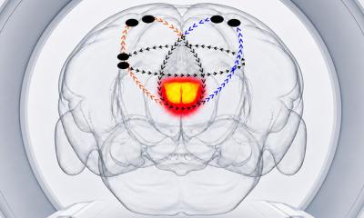
A unique experiment that compares single neuron firing during an activity and again as the activity is freely recalled as a memory shows what the brain looks like during spontaneous recollection.
A unique experiment that compares single neuron firing during an activity and again as the activity is freely recalled as a memory shows what the brain looks like during spontaneous recollection. Hagar Gelbard-Sagiv of the Weizmann Institute of Science in Rehovot, Israel, and Itzhak Fried of the University of California, Los Angeles showed clips of The Simpsons and other television programs to a small group of epilepsy patients with therapeutically implanted brain electrodes, monitoring the activity of single neurons in the brain's hippocampus and related structures as the clips played. As the people freely recalled the shows minutes later, the same neurons increased their firing rate again. The neurons sprang to life even before the people told the researchers that the memory of the show had "come to mind," suggesting a direct link between free recall and neuron "replay" in this portion of the brain.
While you recall that moment that you met your first love, an ever-changing constellation of neurons fires in your brain. Researchers have long hypothesized that each firing pattern should be specific to a particular memory and should be the same regardless of where or when you are having the memory, or what triggered it. A new study in rats has now confirmed this hypothesis. The rats didn't actually fall in love, as far as anyone knows; they were just trained to run through different arms of a maze, switching from one arm to another. They also had to run in a wheel between each trip through the maze. The cage was dark and quiet during this interval, so there were few environmental stimuli for the rats to process. To complete the task successfully, the rats had to remember during the wheel run which arm they had just been in so they could decide which arm to enter next. Eva Pastalkova and colleagues recorded the activity of multiple neurons in the rats' brains as they completed these exercises. The researchers observed the same activity patterns, completed over the same time course, during the maze running and the wheel running. These patterns likely represent the rats' memories of running through the first arm of the maze, the authors say.
This article is adapted from the original press release.
08.09.2008