Epigenomic Analysis
New technology helps personalized medicine
A new technology that will dramatically enhance investigations of epigenomes, the machinery that turns on and off genes and a very prominent field of study in diseases such as stem cell differentiation, inflammation and cancer, is reported in the research journal Nature Methods.
The examination of epigenomes requires mapping DNA interactions with a certain protein in the entire genome. This epigenomic characterization potentially allows medical doctors to create personalized treatment of diseases, by understanding the state of a patient, making the forecast, and tuning the treatment strategy accordingly. However, such tests require a huge number of cells. At one point, the study of in vivo genome-wide protein-DNA interactions and chromatin modifications required approximately 10 million cells for an individual test. This enormous requirement practically ruled out such analysis on patient samples.
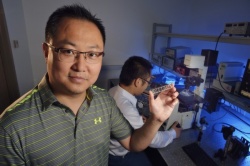
For well more than a decade, Chang Lu, a professor of chemical engineering at Virginia Tech, has worked on the development of tools to effectively analyze living cells with the long-term goal of gaining a better understanding of a range of diseases. In his lab, Lu and his students develop small microfluidic devices with micrometer features for examining molecular events inside cells.
Microfluidics is a branch of science that deals with the performance, control, and treatment of fluids that are constrained in some fashion.
The latest breakthrough comes from Lu’s collaboration with Kai Tan at the University of Iowa, a systems biologist and associate professor of internal medicine. Together, they demonstrated that a technique called microfluidic oscillatory washing based chromatin immunoprecipitation (MOWChIP-Seq) allows analysis of epigenomic modifications using as few as 100 cells. The description of this advance is in the Nature Methods paper.
The National Institutes of Health, along with a seed grant from Virginia Tech’s Institute for Critical Technology and Applied Science, funded this work.
“The use of a packed bed of beads for ChIP allowed us to collect the chromatin fragments with a very high efficiency. At the same time, effective washing for removing undesired molecules and debris guarantees the purity of the collected molecules. These two factors constitute a successful strategy for epigenomic analysis with extremely high sensitivity” Lu said.
The entire MOWChIP process takes about 90 minutes as opposed to many hours that conventional ChIP assays took.
They used their technology to study the epigenomes of hematopoietic stem and progenitor cells isolated from the fetal liver of a mouse in Tan’s lab.
As Tan explained, “Little is known about the dynamics of the epigenome during embryonic hematopoiesis, largely due to the difficulty in isolating sufficient quantities of these cells from developing embyros. This technology is the perfect tool for tackling this problem.”
“Our technology paves the way for studies of epigenomes with extremely low number of cells from animals and from patients,” Lu said. Supported by several NIH grants, the team plans to use this technology to study other epigenomic changes involved in inflammation and cancer in the near future.
Source: Virginia Tech
11.08.2015


