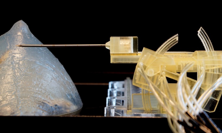
Image credit: Kalle Kataila, Aalto University
News • Larger sample yield
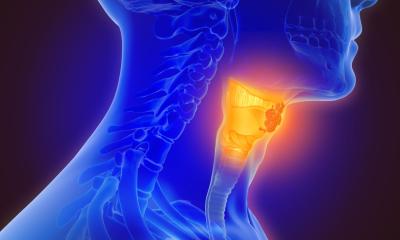
Ultrasonic needle could reshape cancer diagnostics
The minimally invasive approach could allow for in-clinic sampling with significantly more accuracy than fine-needle aspiration. Successfully trialed on patients with salivary gland tumors, it shows strong potential for broader diagnostic use in lymph nodes, and thyroid and breast tumours.
Developed at Aalto University over several years, a new ultrasonic needle for tumour diagnostics has been trialled in collaboration with Helsinki University Hospital (HUS). According to the resulting peer-reviewed study published in the journal European Radiology Experimental, salivary gland tumours could be diagnosed with far greater precision using the innovative needle.
‘With the new method we obtain two to three times more tissue than current needle biopsy techniques,’ says co-lead author Yohann Le Bourlout of Aalto University.
Achieving the most accurate possible diagnosis in the head and neck region before a planned operation helps clinicians tailor the extent of surgery and facilitates patient counselling
Katri Aro
‘Ultrasound travels along the needle’s shaft and is amplified at the tip, vibrating at up to 30,000 times per second. Small fragments of tumour tissue and cells detach and are drawn into the needle by low pressure,’ explains Professor Heikki Nieminen, who led the study.
In tumour diagnostics, a cytological (cell) sample or a tissue sample is essential for both diagnosis and to determine the extent of care needed, says Katri Aro, the other lead author and Chief Physician at HUS. ‘Imaging does not always clearly distinguish between benign and malignant tumours. Achieving the most accurate possible diagnosis in the head and neck region before a planned operation helps clinicians tailor the extent of surgery and facilitates patient counselling,’ Aro says.
Traditionally, tissue is sampled using a core needle biopsy (CNB) or a fine needle aspiration (FNA). CNB is not suitable for all tissues or targets, so FNA is often used because of its thinner needle and broader applicability. Although it causes less pain and carries a lower risk of bleeding, FNA often fails to provide a sufficiently reliable tissue sample and can lead to an inaccurate or inconclusive diagnosis.
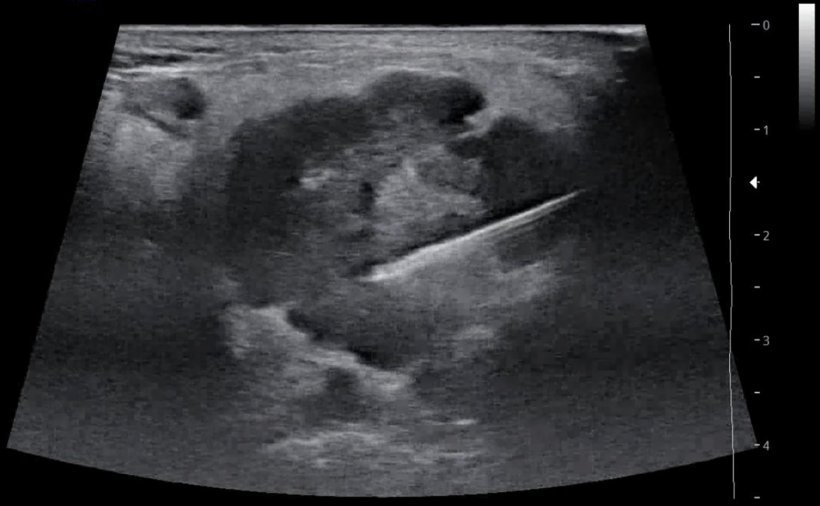
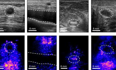
Image source: Naukkarinen M, Le Bourlout Y, Rehell M et al., European Radiology Experimental 2026 (CC BY 4.0)
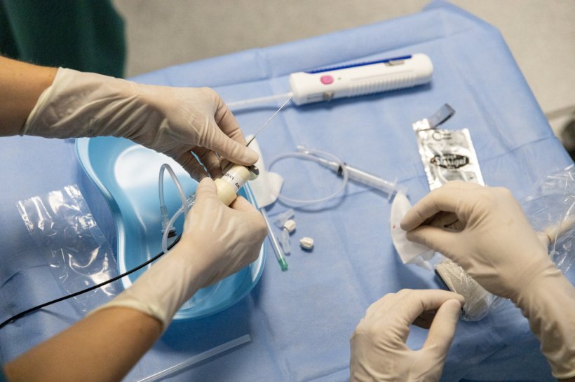
The new, fine ultrasound needle is similar in size to FNA, yet it offers a more precise and larger sample, while being less invasive than a CNB, the researchers explain. ‘The motion at the needle tip helps collect tissue without damaging surrounding structures or causing new complications,’ says Le Bourlout. ‘Yet, the procedure is minimally invasive and can be done at a regular outpatient clinic.’
Salivary gland tumours are rare and diverse, and obtaining samples presents clinical challenges, as the parotid gland lies close to the facial nerve. ‘Until now, it’s often been difficult to obtain a large enough sample to detect the tumour or determine the appropriate treatment, often leading to the need for surgery at the diagnostic stage,’ says Le Bourlout. In combining the strengths of both FNA and CNB the approach offers advantages in the form of avoiding potentially unnecessary, high-risk surgery.
Not only did samples taken with the ultrasound needle contain more tissue than standard FNA samples, but they also represented the tumour more comprehensively than both FNA and core needle biopsies, the researchers say. This is because they include not only loose cells but also tissue architecture, allowing for more precise diagnostics.
According to the team, diagnosis of lymph nodes, thyroid and breast cancers, as well as other tumours, could also be made using the new method. The ultrasonic needle has already been used to successfully sample lymph nodes in ten patients.
Meanwhile, Aalto University, HUS, Turku University Hospital (Tyks), and the Wellbeing Services County of Southwest Finland (Varha) are conducting joint research on thyroid and breast tumours. The study uses tissue samples removed during surgery with a plan to schedule clinical trials in future. ‘We’re excited about exploring the many possibilities,’ says Aro. ‘The clear goal is to improve patient care.’
The cross-disciplinary research team also includes co-lead author HUS doctoral researcher Mira Naukkarinen, HUS pathologist Jetta Kelppe and members of the HUS Diagnostic Center team.
Source: Aalto University
12.05.2026