Interview • Between man and molecule
The hunt for genetic risk factors
Professor Christoph M Friedrich researches the interface between man and molecule. In our interview, Friedrich spoke of the advantages of his work at the institute, the role of medical bioinformatics and its interface with radiology.
Interview: Daniela Zimmermann
Source: Pixabay/PublicDomainPictures/SugarandSkullDesigns (Montage: HiE)
Born in Westphalia, Germany, the professor for biomedical computer science at Dortmund University of Applied Sciences recently took up an additional role at the Institute for Medical Informatics, Biometrics and Epidemiology (IMIBE) in Essen University Hospital. In 2013, the cooperation between the two institutions resulted in a new master’s degree at Dortmund University of Applied Sciences and The University of Duisburg-Essen.
‘Medical bioinformatics,’ explained Christoph Friedrich, ‘not only covers the collection, preparation and utilisation of data in daily medical routine, but also links the analysis of biological specimens and samples, such as gene-analysis, with intelligent evaluation and interpretation systems. Imaging plays an important part here. We are currently studying automated bone age assessments based on X-rays; research based on MRI images and CT scans is also on the agenda.’
So, you are giving radiologists a hand…
‘Generally, research in this field is helpful in two ways. Much radiological work is cumbersome, with quantity and effort ever increasing, which also heightens the chances of overlooking something. AI-supported diagnosis can provide the radiologist with important clues and, in cases of doubt, it can lower the error rate.
‘The second area is the acceleration of working processes. With CT scans in particular, it takes time until the radiologist sees the actual point of interest. Software and anatomic information helps to guide him directly there. This is where structured reporting comes into effect. A specialist should obviously always make the final diagnosis but, thanks to automated analysis, certain checkboxes can be prepopulated, saving effort.’
Structured reporting via AI is not exactly a brand new idea…

‘No, but the research approaches and methods have changed. Up ’til five to six years ago machine learning was carried out with the classic, feature-based approach. This means the objective was to extract and evaluate certain features, such as histograms or grey value distribution from the radiological images. This was comparatively successful, and many commercial products are still working with this method. However, some years ago the deep learning hype began and this overtook the previous approaches within seconds. Convolutional neural networks, i.e. the utilisation of networks for making predictions, make a big difference to simplicity, speed and reliability of study results.
‘Suddenly, data is available in the shortest period of time that would previously have taken months of testing, adapting and validating to achieve. This is the beginning of a journey that’s not finished by far. We are increasingly seeing that the future lies not in deep learning itself but in a skilled combination of deep learning and text mining.’
What are the advantages of this combination?
‘When you link image and textual description, this definitely leads to an improved classification rate and more specific prediction. We have already achieved good results with a similar approach. Using freely available, radiological images and respective captions from medical publications we have trained a network which subsequently predicted accurate captions for the respective images. The programme could make statements such as “you can see a fracture in this specific anatomic region” and to list further key words.’
On this basis, software could support diagnosis. But the patient’s history, i.e. the textual aspect, also plays an important part. Does this not mean that further information such as age, gender, cholesterol levels etc. must also be considered?
‘Most definitely. Electronic capture of data and large variations in its quality remains a big obstacle for implementation. Not all preliminary investigations and specific patient information are electronically stored. Furthermore, the quality of data, laboratory evaluations of radiological images, as well as the semantics of diagnoses, varies significantly, making it very difficult to capture everything in one system. We still have a lot of work ahead of us.’
Apart from diagnostic support you are researching risk predictions. What is the role of medical informatics?
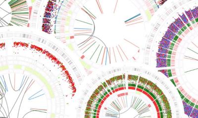
The IMIBE has a large volume of epidemiological and genetic data, which could deliver important medical findings
Christoph M. Friedrich
‘Computer-assisted evaluations help us to filter out important indications for risk factors and their effects, particularly in genetic analysis. An international study, which I was involved in during my time at the Fraunhofer Institute, for instance, had the objective of decoding risk genes that favoured the development of intracranial aneurysms. While a research group in Barcelona analysed image data and developed haemodynamic simulations, in Germany we were searching for risk factors on a genetic basis via text mining – with success. Next to the known associated genetic diseases, such as polycystic kidney disease (PKD), we could identify three new risk factors, so-called SNPs (Single Nucleotide Polymorphisms), i.e. gene mutations.
‘The latest joint research project of the IMIBE and the University of Applied Sciences has a similar approach. We are trying to discover which genes, or gene mutations, push up a patient’s BMI. The IMIBE has a large volume of epidemiological and genetic data, which could deliver important medical findings. As computer scientists we are also interested in larger correlations. One of the theories we would like to validate is that mutations interact and may possibly be addictive or lever each other out. On this level, this is not as much a concrete project as a bioinformatical mission.’
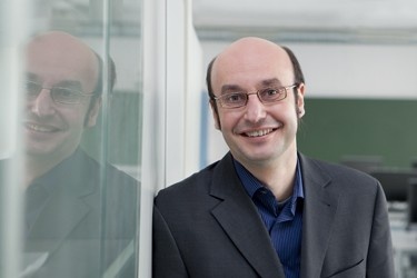
Profile:
Christoph M Friedrich PhD-Eng has been professor for informatics and biomedical informatics at Dortmund University of Applied Sciences since 2013, and recently is also at the Institute for Medical Informatics, Biometrics and Epidemiology (IMIBE) at Essen University Hospital in Germany. His teaching and research focus on medical statistics and machine learning, as well as on image processing and virtual reality. He also temporarily oversees the master’s degree course for medical informatics. After studies in Dortmund he became an IT manager and software developer and, in 2005, became team leader for data-mining in the Department of Bioinformatics at the Fraunhofer Institute for Algorithms and Scientific Computing (SCAI) in Saint Augustin, near Bonn, Germany. A year later he wrote his doctorate thesis at Witten/Herdecke University. In 2010 he was awarded a professorship in mathematics for computer science and applied informatics at Dortmund University of Applied Sciences, before shifting to biomedical informatics.
12.12.2018