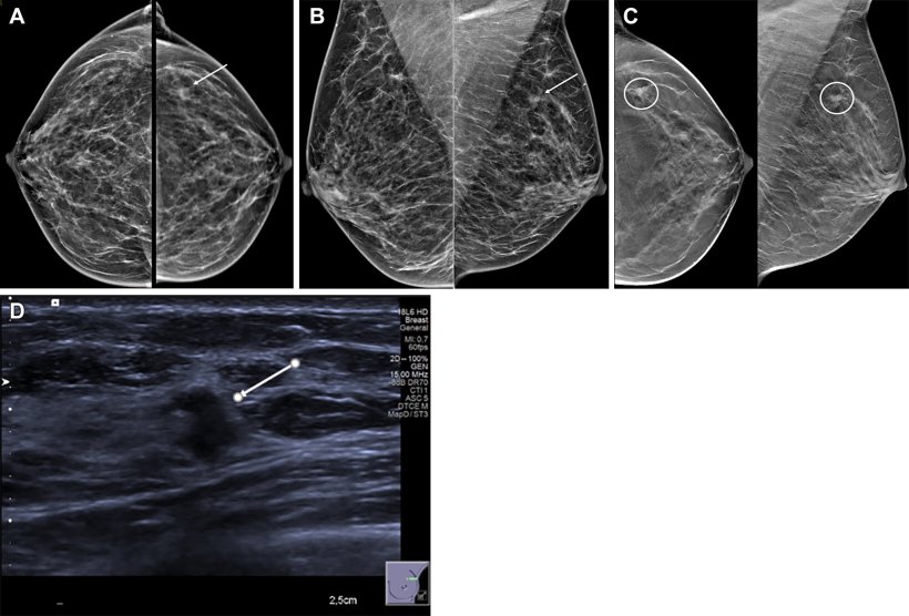
© RSNA
News • Imaging of focal breast complaints
Study confirms ultrasound as effective modality for diagnosing localized breast lumps
Ultrasound is an effective standalone diagnostic method in patients with focal breast complaints, according to a new study. Focal breast complaints can refer to pain, lumps, nipple discharge or other symptoms and conditions confined to a specific area of the breast.
The study was published in Radiology, a journal of the Radiological Society of North America (RSNA).
In women, focal breast complaints are a frequent problem. In the Netherlands, approximately 70,000 women visit radiology departments annually with focal breast complaints. The most common being the presence of lumps or pain. Many women who have focal breast complaints are between the ages of 30 and 50 years. Digital breast tomosynthesis (DBT) followed by targeted ultrasound is the standard diagnostic tool for women 30 years or older with localized breast complaints. While DBT provides an overall image of both breasts, ultrasound can achieve more targeted imaging of a specific area of the breast. The quality of ultrasound images has also vastly improved in recent years.
In a setting with limited resources or an already existing screening program, initial ultrasound might be a better alternative compared to mammography
Linda Appelman
For the new study, researchers set out to investigate the performance of ultrasound alone as an effective diagnostic method in women over the age of 30 who reported localized breast complaints at three hospitals in the Netherlands between September 2017 and June 2019. “The evaluation of breast complaints is a common problem in breast diagnostics,” said study co-author Linda Appelman, M.D., a breast radiologist in the Department of Medical Imaging at Radboud University Medical Center in Nijmegen, the Netherlands. “Ultrasound as the first imaging method may provide clarity with regard to the focal breast complaint.”
Breast lumps or localized breast pain were two of the most common symptoms reported by the 1,961 women included in the study. A total of seven subgroups of focal complaints were recognized. In all patients, targeted ultrasound was evaluated first, followed by DBT. Biopsy was performed after ultrasound, if needed. Since the ultrasounds were conducted first, the radiologists who participated in the study weren’t influenced by the DBT images when interpreting the ultrasounds.
Analysis showed that ultrasound alone led to accurate diagnosis in 1,759 (90%) of the 1,961 patients. Over 80% of the complaints ended up being normal or benign findings, such as cysts. “We found that the diagnostic accuracy of ultrasound is high in women with focal breast complaints,” Dr. Appelman said.
A total of 374 patients had biopsies based on ultrasound findings alone. This resulted in 192 symptomatic breast cancer diagnoses.
Ultrasound of focal breast complaints may be especially beneficial in low- or middle-income countries where ultrasound is more readily available as opposed to DBT. “Our study showed that ultrasound alone can effectively diagnose focal breast complaints in a large majority of women,” Dr. Appelman said. “In a setting with limited resources or an already existing screening program, initial ultrasound might be a better alternative compared to mammography.” The cost effectiveness of ultrasound and the improved patient comfort during the procedure are other factors to consider when expanding its implementation in symptomatic breast cancer diagnosis.
Source: Radiological Society of North America
06.04.2023


