© UZ Leuven
News • Diaphragmatic defect in unborn babies
Smart balloon technique advances fetal surgery
UZ Leuven and its French hospital partner are simplifying fetal surgery with a small balloon that deflates via an MRI scanner
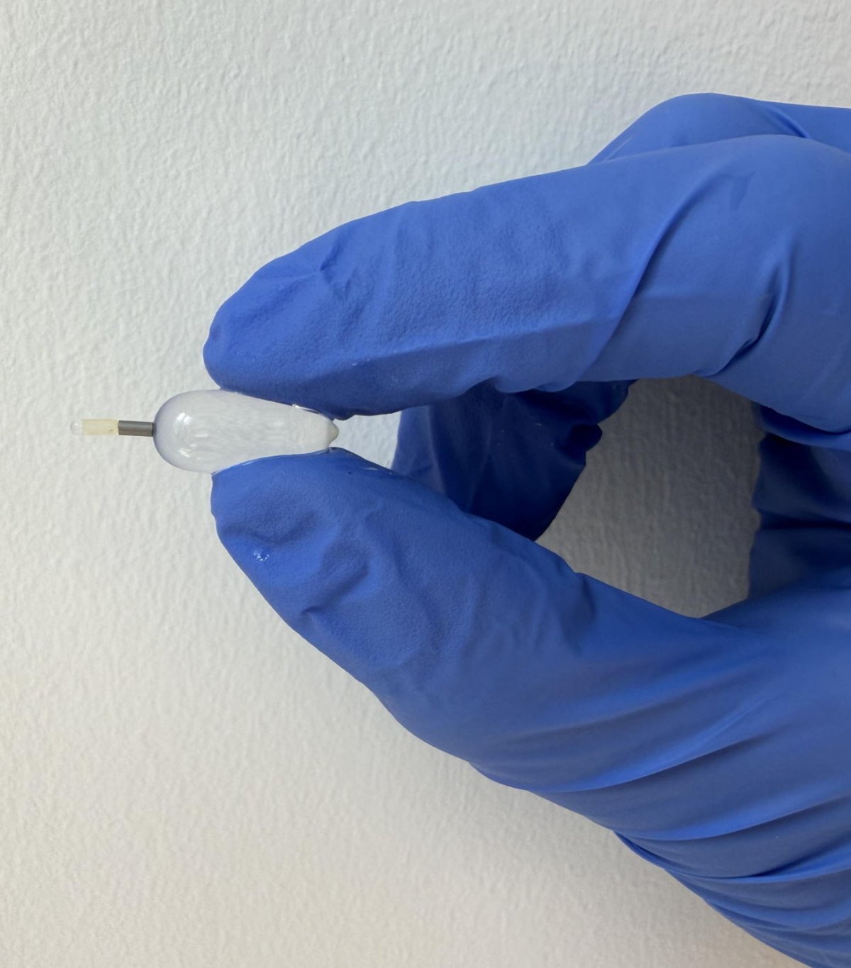
Researchers at UZ Leuven and their hospital partners in France have found a way to avoid second surgery for unborn babies with a severe diaphragmatic defect. To treat this condition, a small balloon is inserted into the fetus’s windpipe, which is normally removed surgically. Thanks to a new ‘smart’ balloon with a magnetic valve, the balloon can now be deflated by exposure to the magnetic field of an MRI scanner. The expectant mother simply walks around the MRI scanner instead of undergoing a further surgical procedure. The results of the study have just been published in the medical journal The Lancet.
This technique makes an existing treatment a lot simpler. We're replacing a surgical procedure by a smart balloon that we can deflate using an MRI scanner
Francesca Russo
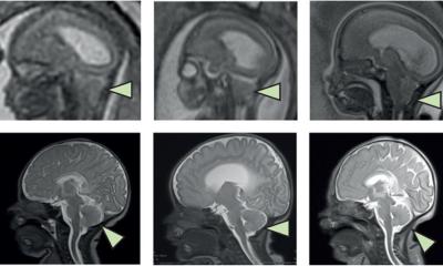
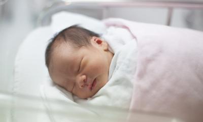
When an unborn baby has a hole in their diaphragm (congenital diaphragmatic hernia or CDH), organs from the abdominal cavity can move into the chest. This leaves the lungs with too little space to develop properly, which can make it difficult for the baby to breathe after birth and may even lead to death.
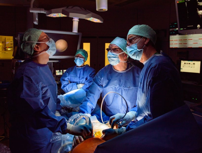
In severe cases of CDH, doctors insert a small balloon into the unborn baby’s windpipe during pregnancy via keyhole surgery. This balloon retains fluid in the lungs, allowing them to grow more effectively. UZ Leuven is renowned worldwide for this FETO technique, which was co-developed in Leuven by an international research team led by UZ Leuven and KU Leuven. The treatment increases the survival rate for severe CDH from around 15% to 40%.
© UZ Leuven
However, the balloon must be removed again before birth. Until now, this has been done via a second procedure, usually around 34 weeks’ gestation. If this is not done in time, the baby will be unable to breathe after birth. This procedure carries further risks, such as premature rupture of the membranes and preterm birth. Furthermore, around one in three pregnant women go into labour spontaneously whilst the balloon is still in place. In such cases, doctors must remove the balloon urgently before the baby is born.
As the procedure on unborn babies is only carried out in specialist centres, expectant mothers often have to stay near the hospital for weeks on end. In 2025, around two-thirds of the patients treated came to UZ Leuven from abroad for FETO treatment. This makes the period particularly difficult for families and, at the same time, requires specialist teams to be on call at all times.

© UZ Leuven
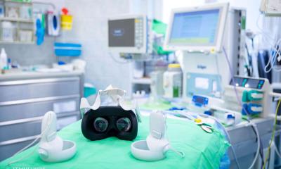
A minor modification to the balloon means that no second surgery is required. The balloon is inserted in the same way, but inside there is a small magnetic bead that closes a valve. To deflate the balloon, the expectant mother now simply walks around an MRI scanner. The magnetic field sets the bead in motion, causing the valve to open and the balloon to deflate automatically. After the procedure, doctors use ultrasound to check that the airway is completely clear. In the study, the balloon was successfully deflated in all cases. Thanks to this new ‘smart’ balloon, part of the follow-up care can take place closer to home in future. This makes the process safer and less burdensome for families and healthcare teams.
Prof. Dr Francesca Russo, fetal surgeon at UZ Leuven, says: “This technique makes an existing treatment a lot simpler. We are replacing surgery by a smart balloon that we can deflate using an MRI scanner. This means fewer risks for the baby, less uncertainty for the parents and care that is easier to plan for the team.”
The study was made possible thanks to collaboration between multidisciplinary teams from Leuven and l’hôpital Antoine Béclère AP-HP, together with the developers of the SMART-TO balloon at the University of Strasbourg.
Source: UZ Leuven
03.07.2026