Small patients, big challenges
Why children are special from a radiologist’s point of view
Children are not small adults. General radiologists are increasingly aware of the very special needs of young patients since frequently they rather than specialized paediatric radiologists perform initial diagnostic imaging and thus play a crucial role in the therapy decision. No mean feat as children may present with very different types of diseases and symptoms than adults, knows Dr. Christoph Heyer, paediatrician and specialist for diagnostic radiology.

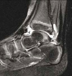
Rheumatic diseases in children for example are a permanent source of confusion simply because we do not expect these pathologies in young patients. “Therefore it is important for radiologists to be aware of the signs indicating rheumatic conditions. Children do not show the typical symptoms that can be seen in adults such as changes and damages in the joints or stiffness. On the contrary: is exactly these advanced stages that we need to prevent. Thus we have to focus on the less obvious signs,” Christoph Heyer points out. These might be joint effusions or changes in the synovial membrane which can be detected in sonography. Imaging combined with physical examinations and lab results allow diagnosis and therapy early enough to stave off irreversible damage.
A second area where paediatric patients present with entirely different symptoms than adults is abdominal tumours. “In children”, Christoph Heyer explains, “it is not liver carcinoma caused by an infection or cirrhosis that is most frequently observed but nephroblastoma, also called Wilms tumour, or neuroblastoma, that is diseases associated with a degeneration of embryonic cells. Both are associated with different symptoms than abdominal tumours in adults.”
Imaging is a crucial component in the diagnosis of abdominal tumours. While ultrasound, in certain cases be complemented by MRI, is the modality of choice CT is usually not required. For radiologists it is important to know these carcinomas, and with regard to neuroblastoma they have to be able to determine the exact size and location of such tumours since they tend to affect the spine.
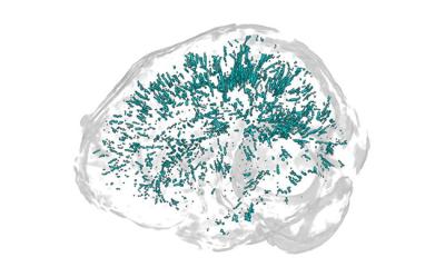
Imaging malformations of the central nervous system is also increasingly important. Firstly, modern prenatal diagnostic imaging can detect many conditions in utero and thus trigger follow-up exams and interventions. Secondly, today MRI is next to EEG the most important diagnostic tool for the diagnosis of seizures. “Seizures usually originate in the brain surface and can cause a number of malformations there. During foetal development neurons migrate to the brain surface where they dock on certain structures. If this does not happen properly seizures or development disorders may occur. This is another area where paediatric diagnostics is quite different from adult diagnostics which focuses on degenerative diseases,” Heyer underlines.
27.11.2012


