MR-Elastography
Technology that measures tissue stiffness quantitatively
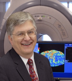
Physicians, over many centuries, have depended on the sense of touch as their hands on method to detect diseases in many body areas. This technique is called palpation. However, though it was known that abnormalities in the stiffness or mechanical environment in tissue may have a profound impact on how many diseases progress, conventional imaging modalities could not display tissue stiffness in a quantitative way. More recently, Professor Richard L Ehman and colleagues at the Mayo Clinic, in Baltimore, USA, have invented a technology for quantitatively imaging tissue stiffness. They call it MR Elastography (MRE).
Already proven in clinical practice at the Mayo Clinic, where MR Elastography has been routinely used since 2007 to detect liver fibrosis – which previously required a biopsy for diagnosis -- the potential of MRE in evaluating diseases of the brain and heart and detecting tumours in the breast, liver, thyroid, and prostate, is now under active investigation. The first commercial MRE application licensed by the Mayo Clinic is being introduced by GE Healthcare, under the name MR-Touch.
When Daniela Zimmermann interviewed Professor Ehman about MRE, the first question was:
How does it work?
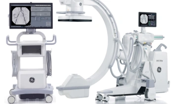
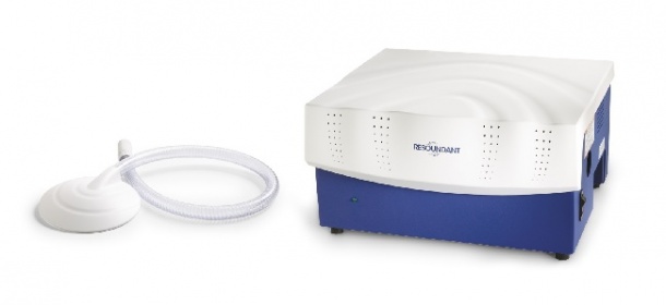
‘We use a conventional MR scanner with additional hardware and software,’ he explained. ‘There are three steps to this technology: First, we generate mechanical waves inside the body by applying vibrations at the surface, using a specially-designed MRI-compatible device. To image the liver with MRE, the device for generating vibrations is a small plastic disc that is placed over the right side of the abdomen. ‘The second step is to create images of the propagating waves inside the body. This is challenging because audio-frequency waves only move the tissue by a few microns. The discovery that made this possible was the invention of a special MRI sequence that can visualise mechanical waves amplitudes as small as a few hundred nanometres in the human body. The acquisition can be very quick, so that we can image the liver with MRE in the time of a single breath-hold.
‘In the third and final step, the MRI system automatically processes the wave images using advanced mathematical algorithms to generate images that quantitatively depict the stiffness of tissue. We call these images elastograms, and they are usually displayed with a colour scale.’
How do acoustic waves reveal stiffness in tissue?
‘We know theoretically that the speed with which waves travel depend on how stiff the medium is. In a stiffer material the wave moves more rapidly, and in a soft material the waves move more slowly. If you take a snapshot of the waves, and if they are all of the same frequency, then the waves that are moving more rapidly will have a longer wavelength and the waves that are moving more slowly will have a shorter wavelength. So by measuring the wavelength at any point inside the material, you can convert that information into an estimate of stiffness. This sounds simple, but it has taken us a number of years to develop reliable mathematical algorithms to convert these images of waves into images of stiffness.’
There are already ultrasound techniques to measure tissue stiffness. What are the advantages in using MRE?
First you have to distinguish between techniques that can measure tissue stiffness quantitatively – like MRE and some newer ultrasound-based techniques – and conventional ultrasound elastography that simply demonstrates the relative deformability of tissue. The latter method has been around for more than a decade, but does not provide quantitative measurements of tissue stiffness. Like MRE, the special quantitative ultrasound-based methods use mechanical “shear” waves to measure the stiffness of tissue. All of these methods require special hardware and software. The equipment for ultrasound elastography is less expensive than an MRI scanner and in the case of the technique called “transient elastography”, it is now in wide clinical use around the world and there is extensive clinical experience.
‘I think that ultrasound-based elastography is ideal for screening applications. MRE appears to have advantages in other areas. In evaluating liver fibrosis, it can be used reliably in patients with obesity or ascites, both of which are common in patients with liver disease. Ultrasound-based elastography of the liver has been reported to fail in up to 25% of patients with obesity. Published studies have shown that MRE has better sensitivity and specificity than ultrasound-based elastography for diagnosing early liver fibrosis. This is probably because MRE provides images depicting tissue stiffness in an entire cross section of the liver, whereas ultrasound-based techniques only provide measurements in spot areas near the edge of the liver.
‘While MRI is a high cost procedure, if MRE is included as part of an already-indicated MRI examination, then incremental cost may be minimal. Finally, it is possible to apply MRE to evaluate tissues deep in the body or structures like brain, which is surrounded by bone and is therefore inaccessible to an ultrasound-based method.
‘We expect that the most important early application of MRE will be in detecting liver fibrosis in patients with chronic liver disease. Nearly 200 million people in the world have chronic hepatitis C infection, and many of these patients will develop progressive liver fibrosis. It is important to identify this subset because treatments are available potentially to prevent progression to end-stage liver disease, which has high mortality. Fatty liver disease is increasingly common in Western countries, and is becoming a major cause of chronic liver disease. For many patients, MRE may offer a safer, more comfortable, and less expensive alternative to biopsy, in this diagnostic setting.
‘We are evaluating the potential of MRE to contribute to the evaluation of heart failure, to asses diffuse brain disease such as Alzheimer’s, and to detect tumours in the breast and prostate.
‘For me, one of the most exciting aspects of our work with MRE is that it is providing access to information about the mechanical properties of tissue in the living body. This type of information has simply not been available in the past. Over the last decade, researchers have begun recognise that the mechanical environment of cells – the stiffness of the extracellular matrix and the forces that are applied – has a profound influence on the biology of the tissue.
‘Recent evidence has suggested that diseases such as organ fibrosis, and even cancer, can be influenced and perhaps even caused by abnormalities in the mechanical environment of tissues. In the past, there has been no way to assess quantitatively the mechanical properties of tissue non-invasively. Now, the availability of MRE and similar techniques is providing access to new and largely unexplored tissue biomarkers that may be important in the new field of predictive medicine and perhaps provide new insights into the cause and treatment of important disease processes.’
05.03.2010