Image credit: Radiological Society of North America (RSNA); source: Zhou Y, Guo X, Lei L et al., Radiology 2026
News • Sharper images, less radiation
Lung cancer management: Photon-counting CT outperforms conventional CT
In a prospective imaging study of 200 adults with lung cancer, photon-counting CT reduced radiation exposure, had fewer adverse reactions and provided higher image quality and better detection of malignant features compared with conventional CT.
Results of the study were published in Radiology, a journal of the Radiological Society of North America (RSNA).
Lung cancer is the leading cause of cancer mortality, accounting for 18.7% of deaths globally. To characterize lung tumors and support personalized therapeutic strategies, doctors use CT imaging. Photon-counting CT is an advanced medical imaging technology that directly counts and measures the energy of individual X-ray photons, unlike conventional CT, which averages them. The result is sharper, higher-resolution images and better tissue differentiation.
We believe photon-counting CT might replace conventional CT in the near future due to its improved imaging quality and the diagnostic confidence it offers
Songwei Yue
“Effective follow-up of cancer patients enhances early detection and survival, with regular imaging identifying recurrence in 60 to 100% of cases,” said Songwei Yue, M.D., chief physician, professor and deputy director of radiology in the Department of Radiology at The First Affiliated Hospital of Zhengzhou University in Henan, China.
However, the cumulative radiation and exposure to contrast media involved in conventional CT imaging pose challenges for lung cancer patients, including the possibility of contrast-induced acute kidney injury. According to Dr. Yue, implementing radiation-reduction strategies for CT is challenging, because it can lead to diagnostic errors.
In the study, researchers compared the benefits of contrast-enhanced chest CT using ultra-high-resolution photon-counting CT versus conventional CT across different populations and body mass indices (BMIs) between June and December 2024. Adverse reactions were documented within one hour after contrast agent injection, and baseline renal function parameters were assessed within 48 hours post-injection.
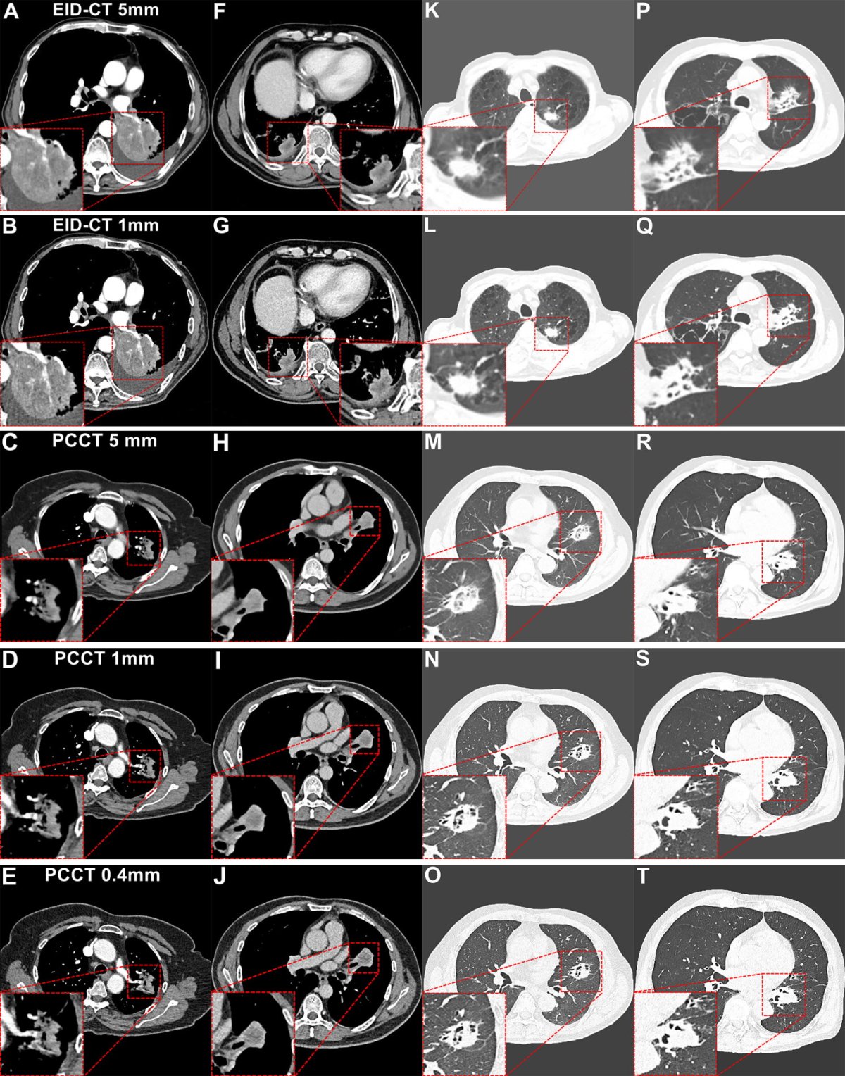
Image credit: Radiological Society of North America (RSNA); source: Zhou Y, Guo X, Lei L et al., Radiology 2026
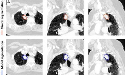
A radiologist experienced in chest imaging performed measurements using a 5-point Likert scale to analyze lesion image quality, malignant radiological features, diagnostic confidence, BMI subgroup and tumor size. Two experienced chest radiologists independently evaluated the noise, anatomical structures, lesion sharpness, structures within enhanced lesions and overall image quality.
Patients were analyzed in subgroups based on lesion size (≤3cm or >3cm) and BMI. Objective image quality and subjective scores were compared among BMI subgroups.
The researchers formed two study groups of 100 individuals, matched on 13 parameters, including weight, age, histological type, T-staging and kidney function. The first group of 100 (61 males, mean age 61) underwent low-dose photon-counting CT, and in the second group (65 males, mean age 61) underwent conventional CT.
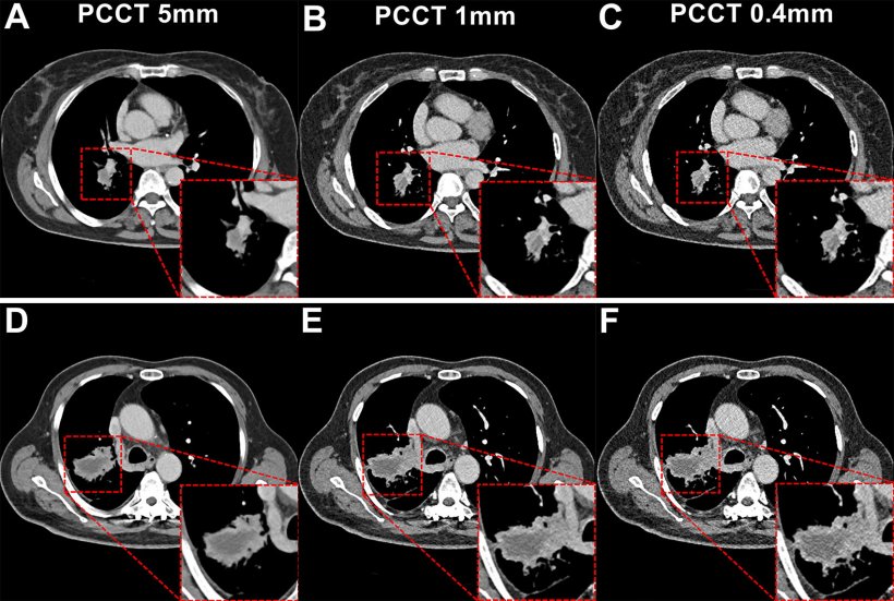
Compared to conventional CT, photon-counting CT reduced radiation and iodine exposure by 66.34% and 26.57%, respectively. Individuals who underwent low-dose ultra-high resolution photon-counting CT also had fewer adverse reactions, including contrast-induced acute kidney injury, compared to conventional CT. At a 0.4 mm section thickness, photon-counting CT improved overall image quality, detection of enhancement-related malignant features, and diagnostic confidence, making it suitable for various BMI and small lesions at the T1 stage.
“Compared with conventional CT, low-dose, ultra-high-resolution photon-counting CT improves the detection of enhancement-related malignant features across varying BMI and tumor sizes,” Dr. Yue said. “It enhanced diagnostic confidence while reducing radiation exposure and contrast media use.”
The researchers said future studies should include longitudinal follow-up scans on the same participants to compare the performance of photon-counting CT in long-term lung cancer monitoring and treatment assessment.
“We believe photon-counting CT might replace conventional CT in the near future due to its improved imaging quality and the diagnostic confidence it offers,” Dr. Yue said.
Source: Radiological Society of North America
07.02.2026