Image source: Oguni T, Izumiya Y, Oda S et al., European Heart Journal - Cardiovascular Imaging 2026 (CC BY 4.0)
News • Combination of LIE and ECV markers
Cardiac CT scans show "invisible" heart risks
Kumamoto University researchers discover that combining two advanced heart imaging techniques can predict life-threatening cardiovascular events with high accuracy.
A routine heart scan might soon do more than just check for clogged arteries; it could act as a crystal ball for a patient's cardiac health. Researchers at Kumamoto University have revealed that by combining two specific markers from a standard cardiac Computed Tomography (CT) scan, they can identify patients at the highest risk for future heart failure and death.
The team published their findings in the European Heart Journal - Cardiovascular Imaging.
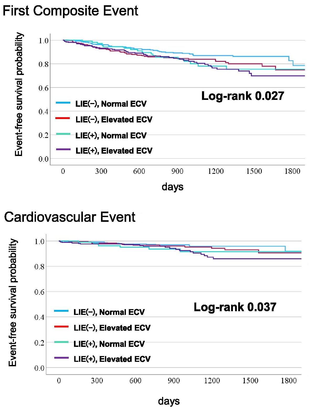
Image source: adapted from: Oguni T, Izumiya Y, Oda S et al., European Heart Journal - Cardiovascular Imaging 2026 (CC BY 4.0)
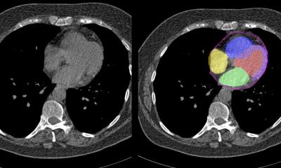
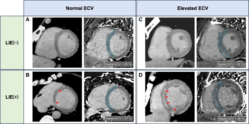
Traditionally, cardiac CT is used to find blockages in the coronary arteries. However, the team led by Professors Yasuhiro Izumiya and Kenichi Tsujita found that the scan can also detect "invisible" damage to the heart muscle itself. By adding a "delayed phase" to the scan, they measured two indicators: Late Iodine Enhancement (LIE), which spots localized scarring, and Extracellular Volume (ECV) fraction, which detects more widespread, subtle damage throughout the heart muscle.
In a study of 1,207 patients followed over an average of 26 months, the researchers discovered that these markers are powerful predictors of a patient’s future. Patients who showed abnormalities in both LIE and ECV were at a significantly higher risk for unplanned hospitalizations or death. "While these two markers provide different types of pathological information, combining them allows us to detect potential heart damage that might otherwise be missed," the researchers noted.
This breakthrough means that a single, non-invasive CT scan can now provide a "synergistic" view of heart health. It offers a faster, more accessible alternative to expensive and time-consuming MRIs, allowing doctors to intervene earlier with life-saving treatments.
As medical technology advances, the heart CT scan is evolving from a simple diagnostic tool into a vital instrument for long-term health management, ensuring that "hidden" risks are brought to light before they become emergencies.
Source: Kumamoto University
24.04.2026