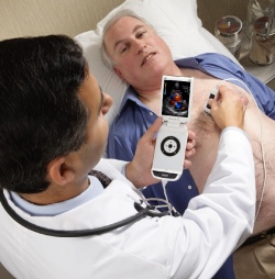
GE’s Vscan takes ultrasound anywhere
Although small, Vscan is a prescription device for ultrasound imaging, measurement and analysis in the clinical applications of abdominal, cardiac (adult and paediatric), urological, foetal/OB, paediatric and thoracic/pleural motion and fluid detection, as well as for patient exams in primary care and special care areas.
George R Sutherland, Professor of Cardiac Imaging at the Department of Cardiological Sciences, St. George’s Hospital Medical School, London, is the first to use the Vscan in the UK. After three weeks he said: ‘I wouldn’t like to be without it. The Vscan provides both high quality moving images of the heart in grey scale and colour Doppler visualisation of blood flow. It’s possible to scan all four chambers of the heart and look at all four cardiac valves and their function in terms of a non-quantitative visual assessment. It’s also possible to identify either important global or regional abnormalities in left ventricular function, as well as to exclude the presence of a pericardial effusion.’
Often asked to triage patients in the emergency room, he added that, with a rapid Vscan, a skilled operator could scan a patient and ‘… either identify the presence of significant cardiac abnormalities or determine that the heart function is normal.’ The device was also used on a number of patients to help detect and monitor fluid aspiration in the pericardial sac.
Vscan is designed like a folding mobile phone, and the interface features a thumb controlled scroll wheel. Handling is intuitive, but Prof Sutherland emphasises that appropriate training and knowledge of ultrasound systems such as this is critical to help in accurate diagnosis. GE Healthcare provides an online portal with training tools and basic clinical applications with sections about imaging technique, anatomy and troubleshooting.
‘A totally portable ultrasound device offers the prospect of decision making from the ICU to the outpatient clinic to ambulatory patients. This is an amazing development,’ Prof. Sutherland concluded.
03.03.2010