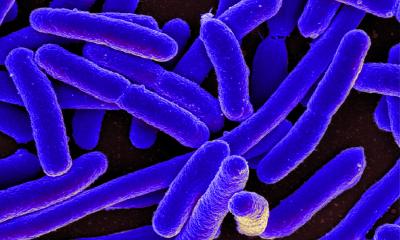
© Dr. Sandor Nietzsche, Electron Microscopy Center
News • Seeing the whole picture
Covid-19 mapping reveals organ distribution and tissue damage
The first known study to measure SARS-CoV-2 viral load in a variety of organs and tissues may aid our understanding of how Covid-19 develops following infection.
Their study of 11 autopsy cases, published in eLife, may contribute to our understanding of how Covid-19 develops in the body following infection. More than 24 million SARS-CoV-2 infections have been reported to date, and the number of deaths attributed to Covid-19 has exceeded 828,000 worldwide. Covid-19 occurs with varying degrees of severity. While most patients have mild symptoms, some experience more severe symptoms and may need to be hospitalised. A minority of those in hospital may enter a critical condition, with respiratory failure, blood vessel complications, or multiple organ dysfunction.
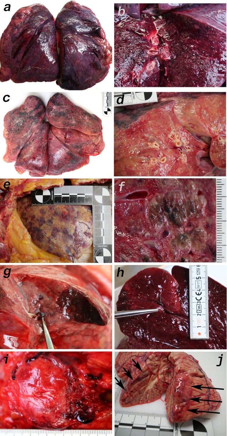
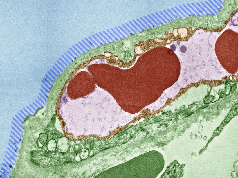
Image source: Deinhardt-Emmer et al., eLife 2021 (CC BY 4.0)
“Clinical observations suggest that Covid-19 is a systemic disease, meaning that it affects the entire body rather than just a single organ such as the lungs,” explains co-first author Stefanie Deinhardt-Emmer, Resident in Medical Microbiology, Jena University Hospital, Jena, Germany. “But we don’t currently have a clear understanding of disease development in humans and other organisms, due to the lack of appropriate experimental models. Investigating the viral distribution of SARS-CoV-2 within the human body and how this relates to tissue damage would help us address this gap.” To do this, Deinhardt-Emmer and colleagues studied 11 autopsy cases of patients with Covid-19. They performed the autopsies at the early postmortem stage to minimise bias due to the degradation of tissues and viral ribonucleic acid (RNA – a molecule similar to DNA).
Their analysis revealed high viral loads in most of the patients’ lungs, which had caused significant damage to those organs. Using an imaging technique called transmission electron microscopy, the team also visualised intact viral particles in the lung tissue. “Interestingly, we also detected SARS-CoV-2 RNA throughout various other tissues and organs unrelated to the lungs that did not cause visible tissue damage,” says co-first author Daniel Wittschieber, Senior Forensic Pathologist at Jena University Hospital. The researchers say that this distribution of viral RNA throughout the body supports the idea that our immune system is unable to respond adequately to the virus’ presence in the blood.
“We show that Covid-19 is a systemic disease as determined by the presence of virus RNA, and yet unrelated to tissue damage outside the lungs,” says co-senior author Bettina Löffler, Director of the Institute for Medical Microbiology, Jena University Hospital. “To our knowledge, this study is the only one to date that has measured viral loads in a wide variety of organs and tissues, with more than 60 samples studied per patient.”
“The insights gathered from our work may add to our understanding of how Covid-19 develops in the body following infection,” concludes co-senior author Gita Mall, Head of the Institute of Forensic Medicine, Jena University Hospital.
Source: Jena University Hospital
30.03.2021