Image source: Pich et al., Nature Communications 2021 (CC BY 4.0)
News • DNA damage causes AML
Cancer chemotherapy side-effects on blood cell development
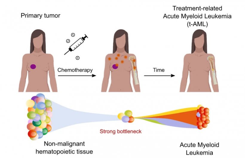
By analysing secondary acute myeloid leukaemias, researchers at the Institute for Research in Biomedicine (IRB) Barcelona have detected mutations caused by platinum-based chemotherapies in cells that were healthy at the time of treatment.
Treatment with chemotherapies influences the development of blood cells, favouring clonal hematopoiesis from cells with pre-existing mutations. The study has been published in the journal Nature Communications.
In the case of platinum-based chemotherapies, the detection of this mutational fingerprint in all AML cells allows us to affirm that the development of treatment-associated AML is subsequent to exposure to these drugs
Núria López-Bigas
Some types of chemotherapy eliminate cancer cells by damaging their DNA. These drugs can also affect healthy cells, where the damage can generate mutations that persist after the end of the treatment.
Researchers at the IRB Barcelona’s Biomedical Genomics Laboratory, led by ICREA researcher Dr. Núria López-Bigas, have identified the "footprints" (in the form of DNA mutations) left by platinum-based chemotherapies in cases of acute myeloid leukaemia (AML) associated with previous chemotherapeutic treatment of a solid tumour. "In this work, we were particularly interested in identifying the mutations caused by chemotherapy in healthy cells, to understand whether the onset of AML is before or after the exposure of patients to chemotherapies," says Dr. Abel González-Pérez, a research associate who has co-led the project with Dr. López-Bigas. "In the case of platinum-based chemotherapies, the detection of this mutational fingerprint in all AML cells allows us to affirm that the development of treatment-associated AML is subsequent to exposure to these drugs," he explains.
Specifically, the researchers were able to identify the "footprints" of platinum-based chemotherapies (cisplatin, oxaliplatin, and carboplatin) in the blood cells of patients with secondary AML. In contrast, despite also being related to cases of secondary AML, therapies based on 5-Fluorouracil or Capecitabine were not found to leave detectable footprints in healthy blood cells, probably because they have a different mechanism of action.
Clonal hematopoiesis is common in advanced ages, and it is the process by which a hematopoietic cell reproduces more efficiently than others, occupying a significant fraction of blood cells over time. Clonal hematopoiesis also occurs as a result of exposure to some chemotherapies. If this process begins after exposure to treatment, mutations in platinum-based chemotherapies would be detectable, as is the case with AML. However, the authors have not identified these mutations, which implies that the onset of clonal hematopoiesis precedes treatment with chemotherapy, which in turn favors its development. "Our study allows us to distinguish whether clonal hematopoiesis had already begun before treatment with chemotherapy and thus establish a temporal relationship," says Dr. Oriol Pich, first author of the study and Alumni of IRB Barcelona, and currently a postdoctoral researcher at the Francis Crick Institute in London.
In 2019, the Biomedical Genomics Laboratory published a paper identifying the "footprints" left in healthy cells by six therapies commonly used to treat cancer. In this regard, they observed that some of these chemotherapies altered DNA between one hundred and a thousand times faster than the processes associated with ageing.
Source: Institute for Research in Biomedicine Barcelona
15.09.2021


