Advanced therapy planning in hepatic surgery
By Dirk Wetzel, medical doctor/engineer at MITI, Klinikum rechts der Isar der TU-Munich
The planning of hepatic surgery of primary and secondary liver tumours is a multimodal process, using modern imaging techniques - mainly contrast enhanced imaging such as CAT and MRI - depending on the patients individual situation as well as on the experience of the medical personnel who are planning the therapy.
New surgical strategies, such as segment oriented and parenchyma-saving resections of tumours and split-liver transplants, created the need for preoperative surgical planning for each individual patient. Due to recent developments in the quality of CAT-technology, PC-based high-performance computer technology and semi-automated software for liver segmentation, virtual and interactive liver surgery planning is now generally available for surgeons. This clinical application is convincingly demonstrated by software developed as a research prototype at MeVis, Bremen, Germany, for hepatic surgery planning.
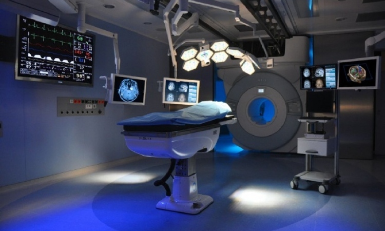
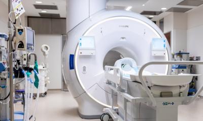
The Somatom Sensation 16 multislice scanner (Siemens Medical solutions, Erlangen) provides highly defined abdominal images, showing the liver, vessel structures and the individual lesion. It was found that the optimal ratio between signal and noise is achieved with a slice thickness of 2 mm for venous contrasted images, and 1 mm for arterial contrasted images. After anonymisation, images are transferred, by a high-speed internet connection, to MeVis for image processing and the creation of a virtual 3D model of the individual patient’s liver anatomy. The software has two components: HepaVision and InterventionPlanner.
Liver and tumour segmentation are performed with a modified live-wire approach, a semi-automated contour finding algorithm. The live-wire contours are interactively determined on a slice about every 10 mm and the contours of intermediate slices are automatically interpolated and optimised. Analysis of the contrast-enhanced vascular systems utilises image filtering, segmentation of the vascular structures with a sectionally increasing algorithm, the determination of main vascular lines, and structural analysis and separation of the resulting vascular trees. This analysis includes the identification of eight major branches of the portal vein, branches of hepatic veins and arterial supply, allowing for a liver segmentation of individual blood supply areas, related to Couinaud’s scheme. Vascular areas and volumes for each vascular region can be calculated separately. A risk analysis is then performed. Based on the determination of vascular structures and their dependant areas, volumetric information can be gained for tumour resections with different safety margins (Fig. 1).
All results are managed by HepaVision and can be displayed in a freely rotating 3D model, or superimposed on the 2D slices. Image analysis takes under an hour, on average.
InterventionPlanner, the second software module, utilises the segmented data to provide interactive generation of resection proposals, with arbitrary safety margins around the tumours and user-defined cutting lines. The volume of remaining liver parenchyma is calculated separately for each resection proposal. (Fig. 2).
The process of oncological liver resection planning is in clinical use at our institution. Virtual resection planning provided valuable information particularly for surgical decision-making in cases of more than one liver metastasis, or of tumours located close to the liver hilus. Precise calculation of the liver volume remaining after different surgical scenarios provides a perspective of potentially curable surgical interventions for an increasing number of patients.
A study of virtual surgical planning vs. the conventional approach is currently underway.
* Researchers: D Wetzel, U Stangl, H Feussner at MITI (www.miti.med.tum.de), and A Schenk and H O Peitgen at MeVis Centre for Medical Diagnostic Systems and Visualisation, Bremen (www.mevis.de)
30.04.2003