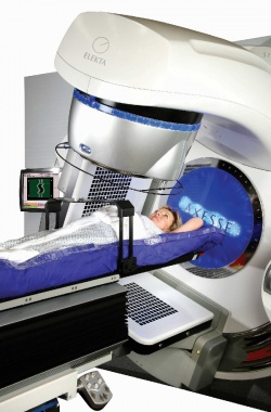
Image guided radiation therapy
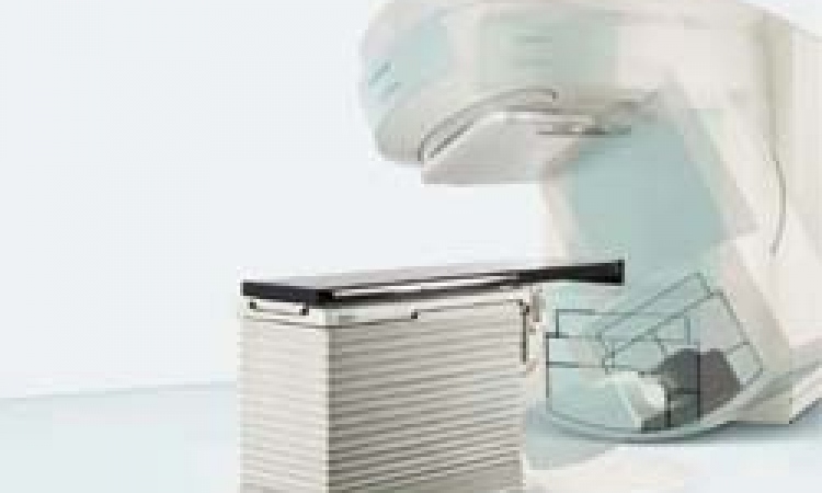
Following the acquisition of an Elekta Axesse system, which provides 3-D image guidance technology for conventional and stereotactic radiation therapy techniques, Helsinki University Central Hospital (HUCH) in Finland reports that, after eight months its image guided radiation therapy (IGRT) of lung, brain, pelvis, head and neck tumours has increased by up to 30 patients daily.
The HUCH clinicians had been using a single IGRT system providing 2-D images, which they found to be not ideal for soft tissue imaging. Mikko Tenhunen PhD, chief physicist at HUCH explained: ‘We wanted a modern treatment system capable of 3-D image guidance and delivery of complex radiation therapy techniques, such as VMAT. We also treat many small tumours, so sophisticated field shaping and patient positioning and immobilisation were critical.’
The team then selected the Elekta Axesse from submitted tenders. ‘Especially for lung and pelvic tumours, 3-D cone beam CT beats a pair of orthogonal 2-D images in terms of image quality and ability to generate images using a low dose,’ Dr Tenhunen said, adding that the chosen equipment has clearly shown the value of 3-D imaging over 2-D imaging, and increased the number of image guided treatments by around 20%.
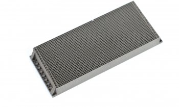
In addition to 3-D image guided localisation at the time of treatment, the Axesse provides ultra-conformal beam shaping with Beam Modulator, the HexaPOD patient positioning system featuring six degrees (x, y, z, roll, pitch, yaw) of remote positional correction, non-invasive patient immobilisation and advanced treatment planning with Monaco.
27.10.2011