Ultrasound guided liver surgery makes tumour removal safer
ALOKA Holding Europe AG, the innovator in ultrasound, is working with the world renowned liver surgeon, Professor Guido Torzilli to explore the clinical benefits of intra-operative ultrasound in hepatic cancer cases.
Ultrasound has one enormous advantage over traditional techniques, such as MRI and CT, since it can be used intra-operatively. The success of this alternative technique for hepatectomies translates into lower mortality rates; mortality rates are up to 5 times lower using ultrasound guided techniques than with traditional techniques.
Intra-operative ultrasound enables surgeons to remove the lesion without damaging too much of the surrounding healthy tissue.”
Without ultrasound guided surgery, 35-50% of surgical cases require a major hepatectomy. However, using Torzilli’s technique means only 4% of surgical cases result in major hepatectomies. Prof. Torzilli says “making the need for major hepatectomies around 10 times less likely is a massive and positive step forward in the care of hepatic cancer patients.” Quality of life and independence are both reduced following a major hepatectomy, so there are further advantages in avoiding major hepatectomies.
There are three types of liver cancer; hepatocellular carcinomas, cholangiocarcinomas and metastatic tumours from colon cancer. All three can be treated with operations to remove the tumours, and, at least for metastatic cancers from colon cancer and cholangiocarcinomas, it is the only treatment which affords a chance of a cure. Prof. Torzilli says “chemotherapy, radiotherapy and other treatments can sometimes keep the cancer under control, but only the association with surgery makes feasible a curative perspective.”
Prof. Torzilli explains the problem: “Although the liver has a remarkable ability to regenerate itself, during operations to remove tumours, it is important to try and remove as little healthy liver tissue as possible, whilst still ensuring all the tumour is removed - to reduce the chance of further growth and relapses. It is this compromise between removing the tumour and sparing the liver tissue itself that I believe is optimised by the use of bespoke intra-operative ultrasound systems.”
Due to the metastatic properties of some forms of hepatic cancers, inoperable relapses can be common when tumours are removed using conventional techniques. In contrast, by operating conservatively in the first instance, a second round of surgery becomes a viable option as less tissue needs removing – meaning liver function can still be retained. Ultrasound is allowing previously unfeasible operations to go ahead, and this is saving lives.
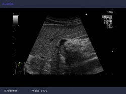
Using a range of probes, Prof. Torzilli is able to give himself the best view at each stage. A convex probe allows him a wide view field to initially explore the area, a T-shaped probe provides resolution for superficial structures, and for guiding surgical maneuvers the Micro-Convex probe.
Prof. Torzilli concluded by saying “Without ultrasound guidance we would never have been able to give these patients such a good chance of overcoming the cancer and retaining good liver function.” This technique is being built back into accepted practice with the training of the new generation of surgeons, and Prof. Torzilli strongly recommends that “all surgeons train in the intra-operative use of ultrasound for hepatectomies.”
06.07.2011