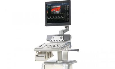
Ultrasound CT for early diagnosis
3D images with 10x higher res
Karlsruhe, Germany - A new type of ultrasound computed tomography (CT) system promising to improve diagnosis significantly is currently being developed at the Research Centre Karlsruhe.

The procedure delivers three-dimensional (3D), reproducible images with a resolution ten times higher than conventional ultrasound images. The centre, part of the Helmholtz Community (www.fzk.de) reports that the first 3D-demonstrator will shortly be available to carry out first examinations on live tissue.
The new system makes it possible to capture even capillary structures with good contrast. In trials, objects such as straws and nylon threads were embedded in gelatine and measured with the tomograph. ‘Even structures of 0.1mm in size with gaps of 0.5mm between them could clearly be recognised,’ said Rainer Stotzka, head of the project. Experts agree that ultrasound CT could soon become the preferred method for early diagnosis, particularly for younger women, because this new system does not share the harmful side effects of X-ray mammography.
Describing the interdisciplinary project, Hartmut Gemmeke, head of the Institute for Data Processing and Electronics (IPE) at the research centre, said: ‘In the development of the ultrasound CT system we have combined innovative concepts from the worlds of sensor technology, microelectronics, high-performance computing and algorithm development.’ The Institute developed a method for the inexpensive production of thousands of miniaturised ultrasound converters required for the production of 3D tissue images. The control logic for the tomograph was also developed and manufactured at the IPE, along with high-performance computers with several gigabytes per second for the processing of large volumes of data.
01.07.2004


