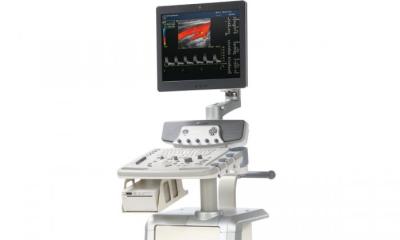
X-ray and ultrasound combo on a mammo platform
A first in medical imaging is still unknown for Kit Vaughan, who is ready to simultaneously scan with X-rays and ultrasound for breast screening. Stay tuned for the results at RSNA 2013, says EH Correspondent John Brosky


It all works fine on paper – and the first clinical trial shows half the system, the X-ray mammography unit, is performing perfectly well. Now, over the next six months, CapeRay of Cape Town, South Africa, will try for the first time to layer an ultrasound scan on top of a simultaneous X-ray acquisition of compressed breasts. The goal is to generate a tightly registered fusion image for the early detection of breast cancer.
The PantoScanner X-ray mammography platform incorporates automated breast ultrasound, which the company believes will greatly improve both sensitivity and specificity for screening of women with dense breast tissue, where 50 percent of cancers are routinely missed.
For radiographic component, the PantoScanner Soteria is a slot scanner where the x-ray tube remains stationary as it emits a fan beam as the collimator, a narrow band detector, moves under the breast.
According to CapeRay CEO Christopher ‘Kit’ Vaughan, the PantoScanner platform requires a more powerful X-ray tube, ‘… but a woman is exposed to much less radiation using slot scanning and we get a higher quality image due to a higher signal to noise ratio that cuts down on the scatter, potentially by 50 percent.’
Just how good the image quality is was not known until October 2012, when CapeRay was authorised to conduct its first clinical trial on 30 women.
A radiology board is currently assessing those images against a predicate device manufactured by GE Healthcare (Waukesha, Wisconsin).
Now comes the simultaneous ultrasound acquisition, the unknown factor. Bound to the detector moving on the PantoScanner, the ultrasound transducer that was added to create the PantoScanner Aceso model must move at 50 millimetres per second, which should be sufficient to acquire the signal.
‘We have the prototype, but we have not yet acquired images,’ Kit Vaughan explained. ‘The effect of the scanning speed on image quality remains unknown, though we have an innovative way to solve the coupling and matching problem.’ On paper, he added, the radiation should not have a detrimental effect on either the ultrasound transducer or image quality.
The PantoScanner Aceso model is an automated breast ultrasound (ABUS) in the same family as the FDA-approved U-Systems Sono V that was acquired in November 2012 by GE Healthcare.
What distinguishes the CapeRay ABUS method, said the CEO, is that where the Sono V requires a woman to lie down for a horizontal image acquisition, Panto uses the traditional standing position with compressed breasts. ‘The issue is co-registration of the X-ray and ultrasound images,’ he pointed out. ‘When a radiologist needs to align two different images in his head, one vertical and compressed with one lying flat, it becomes difficult. Theoretically,’ he added, ‘our system acquires the breast images in the same orientation with the same degree of compression.’
With a goal of launching the combination PantoScanner Aceso model at RSNA 2013, Kit Vaughan says the CapeRay development team is ‘scrambling to get the system ready’.
A clinical trial at the University of Cape Town, already funded by the South Africa Cancer Society, is planned for July 2013.
‘We hope to make a meaningful difference for women in discovering cancers earlier,’ Kit Vaughan explained, ‘and for radiologists to make the job so they get the right diagnosis early enough.’
22.11.2012


