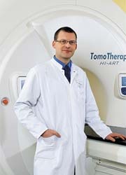
Tomotherapy system targets cancer more precisely
When tumours are situated in difficult locations, e.g. near the brain, lungs, prostate or abdomen, patients treated with conventional radiation equipment often suffer severe side effects, which include bleeding, chronic infections or organ function limitations.

‘This is where tomotherapy opens up new ways of therapy and individually customised treatment,’ explained Professor Robert Krempien, head of the Radiotherapy Department at the Helios Clinic in Berlin-Buch – among the first German hospitals to use tomotherapy against cancer. ‘The new tomotherapy system allows us to treat patients for whom radiotherapy was previously impossible,’ he said.
Worldwide, tomotherapy technology – a combination of linear accelerator and computed tomography (CT) – is considered the most advanced procedure in radiation therapy. Whilst a high resolution CT precisely locates a tumour, before each individual radiation dose, the linear accelerator, rotating around a patient, fires at a tumour from all sides. ‘We protect sensitive organs by radiating the tumour from many different directions and avoiding these sensitive organs,’ Prof. Krempien points out. In addition, the radiation dose that destroys a tumour can be individually customised to the tumour’s density (the number of cancer cells).
The tomotherapy system also enables radiation of several tumours and metastases in one day. Adjusting the radiation dose intensity to the extent, strength and type of tumour presents even better protection of healthy tissue and neighbouring organs. Depending on the tumour, radiation lasts 5-20 minutes.
Additionally, although the location of organs and tumours change continuously, tomotherapy adapts to those changes during radiation. The equipment captures slices indicating the current size and location of a tumour with the integrated CT prior to each radiation, so the beams can precisely reach organs that move.
01.07.2008


