Article • Evolution of technology
The value of AI in breast screening
Although breast cancer (BC) mammography screening enables early detection of breast cancer, mammography presents issues such as variability between the radiology readings and shortage of radiologists. This area of medical imaging is where artificial intelligence (AI) could help make the biggest difference and improve patient outcome, a Netherlands-based researcher told delegates at an April meeting in Spain.
Report: Mélisande Rouger
Millions of mammograms need to be read annually, putting a strain on radiology services. Computer aided diagnosis (CAD) emerged more than 20 years ago for second readings, with markers that highlighted the area of evaluation for further inspection. Overall, the benefit of using CAD is disappointing, Albert Gubern-Mérida, an AI researcher in Nijmegen, explained at the ESR AI Premium meeting. ‘These systems all target the perception level and just help the radiologist double check things that could have been missed. Besides, those algorithms are outdated,’ he pointed out.

AI systems today are trained on millions of images and associated data. ‘Mammography is the best area of application because thousands of mammograms are generated daily around the world and need to be read,’ said Gubern-Mérida, Head of Research and Development at ScreenPoint, a company with commercially available AI software for 2D and 3D mammography.
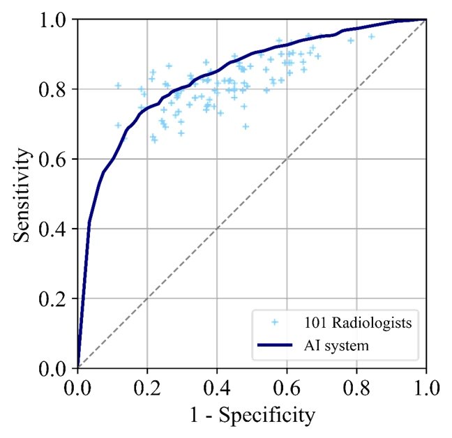
Performance of some current AI systems is at least equal to human performance, research published in the Journal of National Cancer Institute suggests. At Radboud University Medical Centre, researchers independently tested Transpara software vs. 101 radiologists, collecting both multiple centre and multi-vendor data sets. They showed that the algorithm was performing as well as the radiologists’.
Machines will not replace radiologists
Equal performance does not mean machines will eventually replace radiologists. ‘It’s not going to happen, but we need to use this clinically proven, high quality technology to improve the care we give to women,’ Gubern-Mérida emphasised.
There are various ways to apply these technologies, which can improve clinical performance workflow, especially in detection and decision support, recent research suggests. ‘AI tools can highlight things that shouldn’t be missed without interrupting the reader; or, AI results can simply be requested when a second opinion is needed. Studies show that radiologists improve both specificity and sensitivity using these algorithms,’ he said.
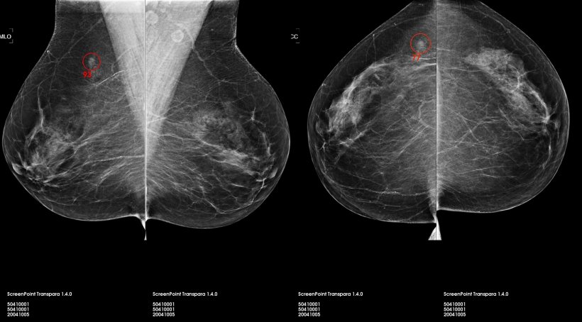
AI may positively impact on workflow by reducing radiologists’ workload. BC mammography screening means reading many images. Second reading improves detection, but this approach might be unsustainable with the introduction of Digital Breast Tomosynthesis, since the reading time increases twofold compared to 2-D mammography. Having an AI system that immediately helps to differentiate exams with and without suspicious lesions could help save a lot of time and trouble. ‘AI systems can produce a score, to detect and determine the risk of cancer presence in an exam. Usually there’s a score from 1-10 or 1-100,’ he explained.
One simple application, possible today, is through the work list, by sorting and labelling exams by their risk of having a cancer – for instance, from the most likely cases to more likely ones, less likely ones, etc. and dividing tasks based on a radiologist’s schedule.
Recommended article
News • Advanced techniques
Breast cancer: how imaging technology will help avoid unnecessary biopsies
Enhancing the diagnosis of breast cancer is the stated goal of a research team at the German Cancer Research Center (DKFZ) in Heidelberg. The scientists have combined an advanced method of diffusion-weighted MR imaging with intelligent image analysis methods to detect malignant changes in tissues. This method may help avoid many control biopsies following suspicious findings from mammography…
AI should make decisions
With an ever-thinning radiology workforce, he suggested, ‘Let AI decide if a second radiologist is needed to read a scan. AI systems are exceptionally good at reading normal scans, so let AI do that.’
Questions from users and ongoing dialogue with PACS providers are essential to ensure they are appropriate and compatible with a customer’s clinical practice. The main questions he said users should ask are: ‘How and where will I use these tools in my workplace?’ and ‘Are these tools clinically relevant for my population and clinical images?’
‘In mammography, we know the images aspect might change, given the different characteristics of devices from different vendors (i.e. detector, angle, processing algorithms, etc.). An algorithm might suffer from these changes.’ Gubern-Mérida has no doubt that BC screening is where AI matters. ‘It’s where women can benefit the most,’ he concluded. ‘AI tools are already there to help radiologists in clinical practice. But, we need more studies to validate the best approach to obtain the best out of it.’
Profile:
Dr Albert Gubern-Mérida gained his joint PhD degree in medical imaging at the Diagnostic Image Analysis Group (DIAG) of the Radboud University Medical Centre in Nijmegen, the Netherlands and the University of Girona in Spain. His research focused on automated analysis of breast MRI, by developing image analysis and machine learning algorithms to detect breast cancer. As a post-doctoral researcher at DIAG he expanded his research to other breast imaging modalities (mammography, digital breast tomosynthesis, and automated 3-D breast ultrasound). In 2016, he joined ScreenPoint Medical, and currently heads Research and Development, focusing on the continuous evolution of AI technology.
02.12.2019