The usefulness of musculoskeletal MRI

A report by Thomas H Magee.

In this sagittal MR image a small radial tear of the meniscus is clearly seen...
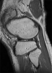
In this sagittal MR image a small radial tear of the meniscus is clearly seen (arrow)
Musculoskeletal MRI has been used for non-invasive diagnosis of joint disease since the 1980’s. In our practice we have seen a continual increase in the utilisation of this imaging modality. Our surgeons have found it very useful in predicting which patients will benefit from surgery as opposed to those who will benefit from physical therapy. Additionally, MRI aids the surgeon in pre-surgical planning, and this can reduce the time required for the patient in the operating room, thus helping to reduce surgical risk. In our experience, musculoskeletal MRI has been useful in excluding patients from surgery. A patient with a negative MRI seldom has positive findings at surgery.

The inherent soft tissue contrast and high spatial resolution of MRI available on high field scanners allows for very accurate diagnoses. Musculoskeletal also has been useful for determining the extent of disease in soft tissue and bony tumours - often not well seen on conventional radiograph or a CT scan. Often, a bone scan can demonstrate increased radiotracer uptake in such cases, but lacks the specificity of MRI. A bone scan will demonstrate increased uptake in trauma, infection or tumour. MRI will demonstrate very specific findings in tumours that help differentiate them from trauma or infection.

MR is useful in demonstrating the presence and extent of infection, which helps guide the right course of action in treating infections. The treating physician is better able to determine whether surgery will be beneficial or whether the infection can be treated with antibiotics only.

Overall we have found MR to be a highly accurate non-invasive diagnostic test for early and specific diagnosis of many types of musculoskeletal disease, and this has significantly improved the treatment of patients with many types of musculoskeletal disease.

02.08.2006

More on the subject:

Related articles

Photo

Sponsored • Diagnostic imaging

MEDTRON AG presents Syriflow® MR: the syringe pump for MR imaging

MEDTRON AG, one of the leading European manufacturers of contrast media injectors and medical technology “Made in Germany,” is expanding its product portfolio with a groundbreaking innovation:…

Photo

News • Biomechanical mapping of tumor growth

New biomarker helps assess glioblastoma aggressiveness

Researchers have developed a new MRI-based method that enables objective quantification of the growth of the most aggressive brain tumours, particularly glioblastoma.

Photo

News • Heart failure diagnostics

AI-enhanced MRI enables single-shot imaging of cardiac cycle

Imaging left ventricular function is challenging for patients who cannot hold their breath for long. A new AI-enhanced MRI technique captures the entire cardiac cycle in a single shot.

Subscribe to Newsletter