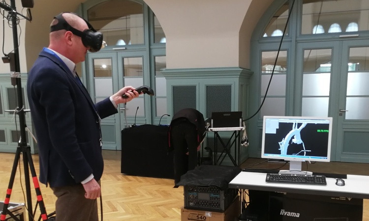
Image source: ESRF; photo: Stef Candé
News • Congenital heart condition
Tetralogy of Fallot: 3D map gives new insights
New 3D map of the heart’s electrical wiring to help patients with congenital heart disease
Researchers from UCL (University College London) and the ESRF (The European Synchrotron) have produced the first three-dimensional map of the heart’s electrical wiring in Tetralogy of Fallot, one of the most common congenital heart problems, revealing anatomical features that may explain why many patients develop heart conduction disorders in this condition. The research, part of the Human Organ Atlas international collaboration, can be used for surgical training and lead to even better outcomes for patients. The research is out in The Journal of Thoracic and Cardiovascular Surgery.
This information certainly is a game-changer reshaping our understanding of the architecture and precise anatomical positioning of the cardiac conduction system in congenital heart disease
Monique Jongbloed
Congenital heart disease affects around 1% of the population worldwide. In many cases, babies must undergo life-saving heart surgery shortly after birth. Although survival rates are now high, many patients develop complications later in life, particularly abnormal heart rhythms or contraction patterns. Surgeons have long known that these problems can arise when the heart’s delicate electrical conduction system, which is invisible during surgery, is disturbed.
Andrew Cook, professor of Cardiac anatomy at UCL and senior author of the study, explains: “I often compare it to renovating a house: you wouldn’t want to start drilling into a wall without knowing where the electrical wires are. The same principle applies to the heart”. Instead, surgeons use ‘anatomical landmarks’ and these have now been revised in the study.
This research is part of the Human Organ Atlas international collaboration. The Atlas is powered by an advanced imaging method called Hierarchical Phase-Contrast Tomography (HiP-CT), developed at the ESRF in Grenoble, France, by an international team led by University College London, UK to visualise anatomy in unprecedented detail.
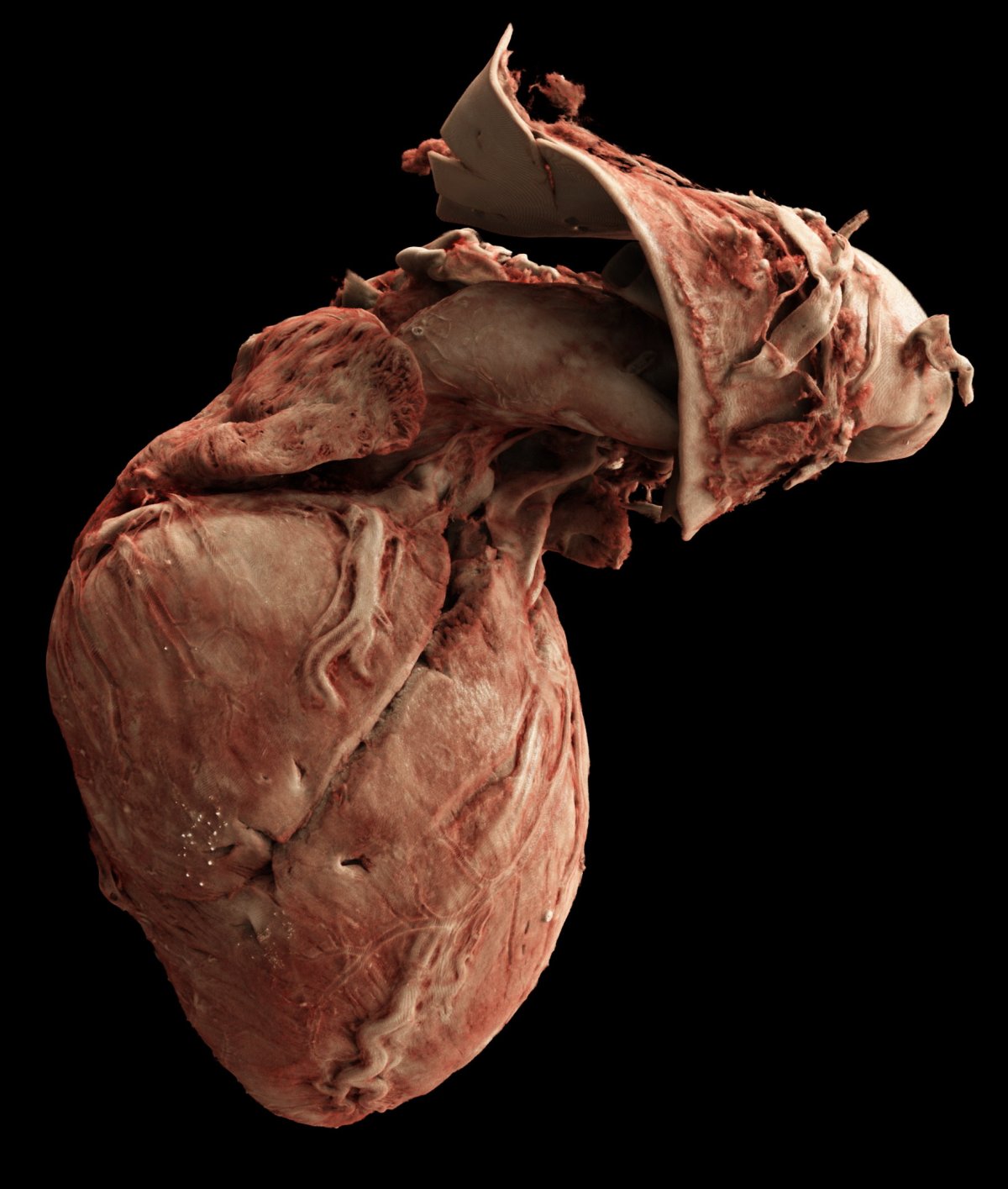
Image source: Joseph Brunet, Cinematic Anatomy (Siemens Healthineers)
HiP-CT uses the ESRF’s Extremely Brilliant Source - a new generation of synchrotron source – which provides an X-ray beam intensity typically up to one million times higher than that used in conventional hospital CT scanners, enabling these organ scans. This allows researchers to scan whole human organs, ex vivo and non-destructively, and then zoom in to near-cellular resolution (down to 2 microns, 50 times thinner than the size of a human hair).
“For more than a century, radiology and histology have offered very different views of the body. The HiP-CT synchrotron technique finally bridges that gap and represents a major step forward in biomedical imaging”, explains Joseph Brunet, researcher from UCL and visiting scientist at the ESRF.
Using scans performed at the ESRF, the researchers examined, non-destructively, 18 whole human heart specimens, with and without the disease. The scans allowed the team to reconstruct in 3D the fine network of specialised fibres that carry electrical signals through the heart, called the cardiac conduction system. For decades, doctors have known where this electrical pathway begins, but how it travels through the heart muscle has remained largely unknown.
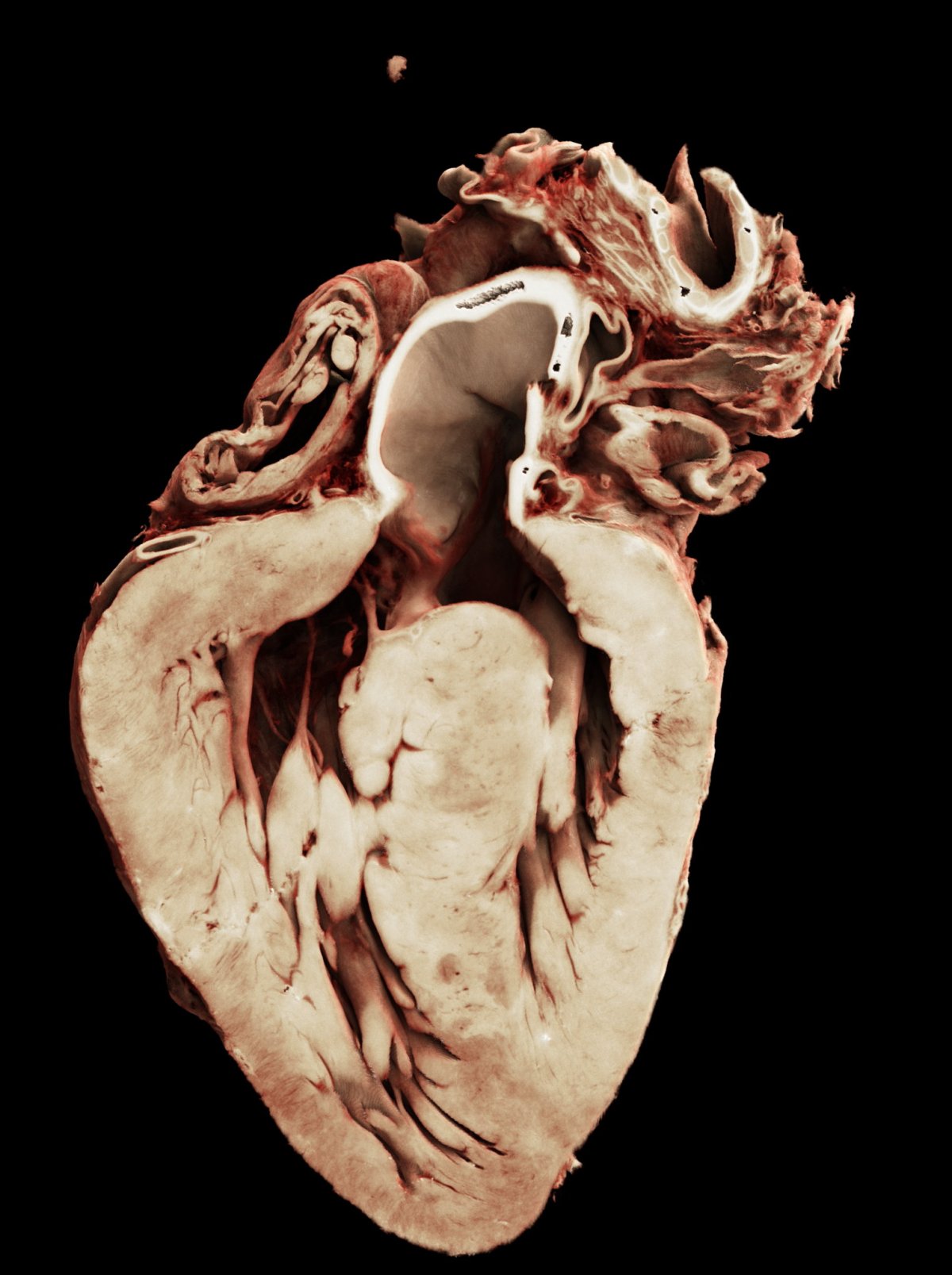
Image source: Joseph Brunet, Cinematic Anatomy (Siemens Healthineers)
The results showed that in tetralogy of Fallot, the electrical wiring of the right ventricle - which is the main chamber affected in the disease - does not run in the usual way as seen in healthy hearts. Instead, the wires are both thinner and spread out across the septum (the wall separating the ventricles) in a way that looks like a piece of fabric draped over a surface.
These findings provide a clearer picture of the anatomical structures that surgeons must navigate when repairing congenital heart defects. “We operate in an extremely challenging environment, often in life-and-death situations and on very small hearts. Any new knowledge about the heart’s anatomy can help us refine our surgical techniques and, ultimately, improve both short and long-term outcomes for these patients”, says Adrian Crucean, specialty doctor in congenital heart surgery at Birmingham Children’s and Queen Elizabeth Hospitals (UK).
By identifying where the electrical wiring runs within the heart muscle, the research could help surgeons avoid damaging it during procedures and potentially reduce the risk of postoperative electrical disorders. “We see many adult patients who have had successful operations during childhood, but later on develop complications such as abnormal heart rhythm. We really want to give them the best quality of life we can, and this information certainly is a game-changer reshaping our understanding of the architecture and precise anatomical positioning of the cardiac conduction system in congenital heart disease”, explains Monique Jongbloed, adult congenital cardiologist at Leiden University Medical Center and part of the EuReCCA consortium.
The researchers also developed advanced computational tools to analyse the large imaging datasets and visualise the results in immersive 3D environments, enabling detailed study of the heart’s wiring. “We have shown that this information can be brought into virtual reality and printed in 3D, so it can be used for training surgeons. It’s never been possible to see the conduction system in this way before”, explains Vaishnavi Sabarigirivasan, PhD student at UCL and corresponding author of the publication.
Recommended article
Article • Cinematic Rendering in surgery
3D visualisation is like ‘body cinema’
What if surgeons could explore the body like a movie set – every vessel, tissue layer, and tumour rendered in photorealistic 3D before the first incision? Siemens Healthineers' Cinematic Rendering does exactly that. The technology transforms CT and MRI scans into strikingly lifelike 3D visualisations – offering surgeons a clearer spatial understanding of individual anatomy.
This research takes place in the framework of the Human Organ Atlas collaboration. The Human Organ Atlas brings together some of the most detailed 3D images of human organs ever produced. It is now a new open-access 3D portal that allows users to explore intact human organs in unprecedented detail — from the whole organ down to individual cells locally. “These data are the result of several years of development of the HiP-CT technique. It was initially developed at the ESRF during the Covid-19 pandemic to study human lungs. In few years, the data quality and acquisition speed dramatically increased, making possible to scan enough organs to perform relevant studies on important pathologies”, says Paul Tafforeau, ESRF scientist and pioneer of this imaging technique used to create the Human Organ Atlas.
“To create the Human Organ Atlas, we brought together scientists and medics from nine institutes worldwide. This group is continuing to expand, helping gain new insights into diseases from osteoarthritis to heart disease and changing how we learn about the human body,” says Peter Lee, Professor at UCL Department of Mechanical Engineering, principal investigator of the HOA beamtime.
“The Human Organ Atlas shows what team science can achieve at its best - we went into this project wanting this data to be used by others and to help further the understanding of human physiology. The Human Organ Atlas is an incredible resource that will continue to grow. I am personally hugely excited to see how the AI community use the Human Organ Atlas in AI foundation models,” says Claire Walsh, Associated Professor at UCL Department of Mechanical Engineering, Director of the Human Organ Atlas Hub.
The members of the EuReCCA consortium are scientists and clinicians from London, Paris, Leiden and Birmingham with specific expertise in the structural architecture of the heart. They are expanding their research to other forms of congenital heart disease such as ‘single ventricle disease’ and invite partners from Europe and beyond to collaborate. Their aims are ‘deep-phenotyping’ of congenital and acquired heart disease; the fast translation of this type of research into clinical practice and surgical training; and the generation of open-access data to allow scientists to create ‘digital twins’ of the heart’s function and electrical conduction.
Source: European Synchrotron Radiation Facility
02.06.2026