Image source: Microfludics Cluster UPV/EHU
News • Bioelectronics for cancer screening
Smart devices to capture and release tumour cells
A group at the University of the Basque Country (UPV/EHU) is proposing the development of low-cost universal platforms for early cancer screening.
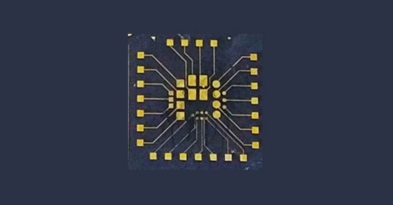
In the book entitled Microfluidic Systems for Cancer Diagnosis, the UPV/EHU’s Microfluidics Cluster group describes the process for building a bioelectronic device consisting of gold electrodes coated with a smart polymer capable of capturing and releasing cells in a non-invasive, controllable way while monitoring the processes using conventional electrical measurements. These are the first steps towards developing universal platforms for early cancer screening.
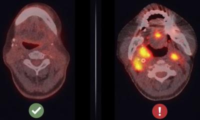
Metastasis is the leading cause of death in cancer, occurring when a cell leaves the primary tumour, passes into the bloodstream and lymphatic system and reaches distant organs. Non-invasive collection of these circulating tumour cells is essential for the study of cell biology, the diagnosis and prognosis in cancer research, and drug development. As a general rule, the concentration of cancer cells found in blood is very small with respect to other cell types, and traditional methods of collecting them in a viable way are laborious.
“We wanted to come up with a device capable of concentrating cancer cells in order to detect their concentration,” explained Janire Sáez, Ikerbasque research professor in the UPV/EHU’s Microfluidics Cluster Group. The biosensors (devices for measuring biological or chemical parameters containing a component of a biological nature) developed so far for this purpose damage cells during the capture and release processes, and so the Microfluidics Cluster group has combined smart materials with the area of bioelectronics (which is about applying carbon-based semiconductors) so that the capture and release of cancer cells can be measured.
Through this device, cancer cells are being selectively reconcentrated in order to detect their concentration
Janire Sáez
The procedure has been detailed in a chapter of the book Microfluidic Systems for Cancer Diagnosis, geared towards the scientific community and which explores the latest advances in microfluidic technologies for cancer diagnosis and monitoring. The book is an ideal guide to the laboratory construction of microfluidic devices specifically developed for cancer diagnostics and to promoting the development of new and improved diagnostic devices. As the Ikerbasque research professor explained, “we show a bioelectronic device consisting of microfabricated gold electrodes coated with a smart polymer (which reacts to temperature changes) and which enables the non-invasive capture and release of circulating tumour cells to be made, and the simultaneous electrical and optical monitoring of the entire process to be conducted”.
“Our tests were performed in culture media; we did not use actual patient samples, but commercial cells sustained in cell culture. We confirmed that our device enabled us to capture and release the cells,” explained the researcher. Now, they are working to tailor the polymer specifically to different cell types. The device “is the outcome of collaboration with a group at the University of Cambridge, with whom we continue to collaborate, and where the device is currently being applied to samples from oesophageal cancer patients. Through this device, cancer cells are being selectively reconcentrated in order to detect their concentration”, said Sáez.
The researcher pointed out that these are “the first steps towards developing platforms for cancer screening. This could be a good step forward because they generally involve low-cost technologies and can be mass-produced. The idea is to use this type of technology for early cancer diagnosis”.
Right now, the Microfluidics Cluster group is focusing its studies on the development of “micrometric structures for bioelectronic devices of this type. We are also developing 3D systems to create 'organ-on-a-chip' systems (biomimetic systems that simulate organs in the human body)”, she concluded.
Source: University of the Basque Country
12.09.2023