Secret disclosed about self-healing embryos
The Weizmann Institute of Science in Rehovot, Israel, is one of the world's top-ranking multidisciplinary research institutions. Now a molecular genetics research team around Prof Naama Barkai found out that an inhibitor molecule channels the morphogenic substances within the injured embryo so that new growing tissues and organs are developing in the right proportions.

From left to right: Prof Naama Barkai, Benny Shilo and Danny Ben-Zvi
Profs. Naama Barkai, Benny Shilo and research student Danny Ben-Zvi of the Science’s Molecular Genetics Department, together with Prof. Abraham Fainsod of the Hebrew University-Hadassah School of Medicine, Jerusalem, have now discovered the mechanisms that enable a half embryo to maintain its tissues and organs in the correct proportions.
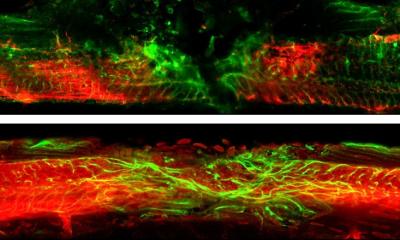
More than 80 years have passed since the German scientist Hans Spemann conducted his famous experiment that laid the foundations for the field of embryonic development. After dividing a salamander embryo in half, Spemann noticed that one half – specifically, the half that gives rise to the salamander’s 'belly' starts to wither away. However, the other 'back' half that develops into its head, brain and spinal cord, continues to grow, regenerating the missing ventral half and develops into a complete, though be it smaller, fully functional embryo. Spemann then conducted another experiment, where this time, he removed a few cells from the back half of one embryo and transplanted them into the belly half of a different embryo. To his surprise, this gave rise to a Siamese twin embryo where an extra head was generated from the transplanted cells. Moreover, although the resulting embryo was smaller than normal, all its tissues and organs developed in the right proportions irrespective of its size, and functioned properly. For this work, Spemann received the Nobel Prize in Physiology or Medicine in 1935.
Previous studies have shown that the growth and development of cells and organs within the embryo is somehow linked to a special group of substances called morphogens. These morphogens are produced in one particular area within the embryo and then spread throughout the entire embryo in varying concentrations. Scientists then began to realize that the fate of embryo cells, that is to say, the type of tissue and organ they are eventually going to develop into, is determined by the concentration of morphogen that they come into contact with. But this information does not answer the specific question as to how proportion is maintained between organs.
The idea for the present research came about when Weizmann Institute scientist Prof. Naama Barkai and her colleagues developed a mathematical model to describe interactions that occur within genetic networks of an embryo.
The data ascertained from this model suggest that the way morphogens spread throughout the embryo in different concentrations is different than previously thought. The team predicts that an inhibitor molecule, which is secreted from a localized source at one side of the embryo and can bind the morphogen, acts as a type of ferry that 'shuttles' the morphogen to the other side. Therefore, the mathematical model suggests that it is the interactions between the two substances that enable the embryo to keep the relative proportion between organs constant, irrespective of its size. Indeed, these predictions have been validated by experiments conducted on frog embryos by the research team.
The importance of the role of these morphogenic substances, as well as their mechanism of action, is evident by the fact that they have been conserved throughout evolution, where different variants can be found to exist in species ranging from worms to fruit flies and up to higher species including humans. Therefore, understanding the processes that govern embryonic cell development could have many implications. For example, it may lead, in the future, to scientists being able to repair injured tissues.
03.07.2008
More on the subject: