Schizophrenia
By Karen Dente
Imaging water molecules in the brain correlates structural and functional deficits
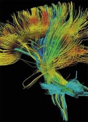
Within the sphere of molecular imaging, Diffusion Tensor Imaging (DTI) provides information about the diffusion of water molecules in biological tissues. Research published in the American Journal of Psychiatry (March 2007) by Dr David I Leitman and colleagues, looked into the neuro-anatomical abnormalities associated with schizophrenia using DTI. The information gleaned from this study allowed them to evaluate the relationship between brain structure and mental function in schizophrenia.
How does DTI work? It gives information about the properties of water diffusion in brain tissue and the integrity of white matter, which are the long axons that radiate from the body of the brain cell, or neuron. The neuronal bodies comprise the grey matter, with axonal tracts making up the white matter covered by fatty myelin sheaths.
This kind of imaging gives information about the integrity of white matter by measuring the diffusion of water molecules. Normally, there is more diffusion of water in the direction parallel to axonal tracts than perpendicular to these, which means it is highly anisotropic. A reduction in this anisotropy of water diffusion, or directionality, is thought to reflect a diminished integrity of white matter, which can be seen in certain diseases affecting the brain.
One theory behind social communicatory dysfunction in schizophrenia is that there are underlying structural changes in white matter integrity on the low-level of visual and auditory sensory input radiations or ‘wiring tracts’ to brain regions responsible for relaying these on to higher processing levels.
A combined look using this imaging with other tests to see whether individuals with schizophrenia can process physical auditory stimuli, such as certain changes in voice, like inflection, or changes in tone (also known as prosody), have shown a correlation of brain region deficits with functional deficits.
Dr Dan Javitt, a co-investigator, says of this study that it was the first time that anybody had correlated structural changes using brain-imaging studies and linked these with functional deficits in emotional processing, the latter of which has been demonstrated in other studies.
Use of such imaging can help focus on the areas of the brain where the problems are anatomically localised and may lead to early detection and intervention by demonstrating a reduction in white matter tracts in people with schizophrenia before the synapses break down. According to the neuronal disconnection model of the disorder, it is thought that the synapses involving the neurotransmitter glutamate seem to be losing their tightness, resulting in a reduction in the number of synapses. This is known from post-mortem studies.
A result of normal synaptic ‘pruning’ during adolescence taken a step too far – some scientists surmise.
New studies are underway in the United States to prevent the disorder from becoming chronic in young adolescents by early intervention to stop cognitive deficits seen in some patients from progressing. Along with other tests, imaging will play a seminal role in early diagnosis.
30.10.2007