Refinements and advancements galore
In essence an upgraded flagship ultrasound scanner is a new system
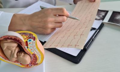
The Aplio, Toshiba’s flagship ultrasound scanner, has been extensively advanced and essentially turned into a new system. The highly innovative Japanese company in imaging diagnostics has refined the successful Aplio series of scanners and introduced it internally in Frankfurt at the beginning of October.
International clinicians who were given the opportunity to test the equipment before the launch were enthusiastic about the precise imaging, compact and smart design and the traditionally high processing quality. Gastroenterologist and liver specialist Dr Horst Kinkel, Assistant Director of the Medical Clinic II at Düren Hospital, confirmed: ‘Compared to the previous version of the Aplio, the technology has become more sophisticated and powerful.’
With its innovative technologies, unique applications and a newly developed hardware and software architecture, Aplio raises the bar in ultrasound diagnostics. Precision imaging, differential imaging and other technologies based on high-density beamforming have advanced ultrasound imaging. The intuitive and flexible operational concept iStyle simplifies complex examination processes.
QuickScan facilitates fast patient-specific optimisation in the B-image and Doppler.
The 3-D FlyThru Tool has been further perfected and is now probably a competitor for hysteroscopy in gynaecological examinations.
The real-time fusion of CT and MRI images with ultrasound scans has become faster and easier.
The new advanced real-time applications in turn expand the diagnostic spectrum and provide additional information to the user through parametric visualisation of tissue changes. The new shear-wave elastography function supports the localisation and assessment of stiffness of tissue inside the body with high precision, sensitivity and reproducibility. The different grades of elasticity can be visualised with colour coding in parametric images and, importantly, can also be quantified – an exciting technology to assess liver diseases such as fibrosis.
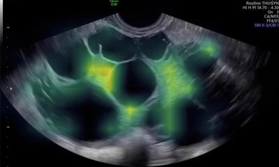
SMI with and without contrast media
With the help of special new algorithms the platinum version of the Aplio can show even the smallest amounts of contrast media selectively in the contrast-imaging mode, making it possible to visualise the finest vascular structures and subtle blood perfusion in lesions and organs. The new Superb Micro Vascular Imaging (SMI) on the other hand shows the blood flow in the vessels right down to sub-millimetre levels – without the addition of contrast media. With the addition of contrast media even in the smallest dose the resolution and sensitivity for blood flow is increased to levels previously unattainable. This facilitates new opportunities for diagnosis in different clinical applications.
SMI visualises organ perfusion right down to the surface layers or the complete vascular architecture in lymph nodes and indeterminate space-occupying lesions. ‘This means that, in the future, it will probably be possible to use contrast media more efficiently,’ Kinkel hopes. This obviously has financial but above all timesaving advantages: ‘All I have to do to utilise SMI is press a button. However, the administration of contrast media, necessitates the availability of syringes and emergency equipment.’ he adds. ‘The scanner shows me at the touch of a button whether there is something there or not – this is a big advantage in this technology. Therefore I rate this innovation so highly – it makes my daily clinical routine easier.’
Detail resolution provides diagnostic safety
‘SMI is a new Doppler-technology with the advantage that tissue artefacts, which normally interfere with the colour Doppler image and make it unusable, can be extremely efficiently suppressed,’ he explains.
‘SMI facilitates high detail resolution and a high frame rate. This provides the doctor with improved morphological understanding and therefore more assurance and safety in making a diagnosis.
‘The complex SMI algorithms make it possible to analyse echo signals and to achieve a distinct differentiation between blood flow and tissue artefact.
‘At the same time they make it possible to recognise the correlations of tissue structures more clearly, which helps, for example, in the diagnosis of patients with abscesses resulting from inflammatory bowel diseases, for example diverticulitis.
‘Now I can determine far more precisely whether there is oedematous tissue, or still healthy, perisigmoid tissue without the presence of abscesses; or I can recognise tissue that’s only of low echogenicity in the normal image despite being well perfused. This,’ Kinkel concludes, has a large impact on my treatment of patients.’
Profile:
After gaining his medical degree and doctorate at the University of Cologne in 1994, Horst Kinkel began his career at Düren Hospital. He qualified as a specialist for internal medicine in 2001 and was awarded the Instructor Level II of the German Society of Ultrasound in Medicine in 2004. At the end of 2008 he became Assistant Director of Medical Clinic II at Düren Hospital, where his focus is on contrast ultrasound, interventional ultrasound, haepatology and chronic inflammatory bowel disease. Dr Kinkel is a member of numerous scientific societies, co-author of textbooks and of an interactive instructional CD.
31.10.2014