Paediatric radiology
Small patients, big needs
Children are not small adults. As well as special technical requirements, their treatment needs particular handling by the radiology team.
In Europe, qualifications to become a paediatric radiologist are not too consistent. According to the report Radiological Training Programmes in Europe, produced by the European Association of Radiology (EAR), countries such as Norway, Poland, Portugal, Italy and Greece do not require radiologists to train for paediatric radiology. However, Germany, Ireland and Romania require three-year paediatric radiology training, whilst some other countries require five years of training.
Due to special indications and particularities of child anatomy, paediatric radiology calls for considerable experience as well as a trained, diagnostic eye. The internet also offers individual training offers and opportunities for experts to exchange ideas, reading lists and research material to help with case studies. For example, the website www.pedrad.info, accredited by the American Roentgen Ray Society (ARRS) and the Radiological Society of North America (RSNA) offers comprehensive training and links, as well as a list of paediatric radiologists working in hospitals and clinics in Europe and the US.
Apart from specialist medical knowledge, dealing with young patients calls for empathy and a way of explaining things that is appropriate for children. Quite often the children’s fear of examinations that involve large equipment, such as CT or MRI, is bigger than the pain they might be experiencing. Children’s books, such as comics about a little heroine MAXX (e.g. ‘That’s me – MAXX’) published by Schering, explain the function of CT and MRI examinations in funny stories, so children can find out what happens in the big tube, ‘who’ takes the pictures and ‘why’ lying still is so important. Parents also need to receive full information, so that they can adequately prepare a child for examination, as well as have reassurance themselves, particularly concerns about radiation exposure that might be unfounded.
Technically there have been rapid developments in recent years that offer many diagnostic and therapeutic chances for children. For example, the LightSpeed Volume Computer Tomograph (VCT), made by GE Healthcare, facilitates the non-invasive diagnosis of cardiac defects in babies and children, e.g. congenital. Due to the detailed 3-D depiction of the heart and coronaries, plus fast image acquisition in less than five heartbeats, this method provides a real alternative to catheter examination.
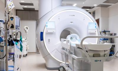
The Panorama 1.0 T MRI scanner, from Philips, offers a particularly child-friendly solution for MR examinations, because the completely open system counteracts claustrophobia in patients and, even more important, children can remain in close contact with their parents during a procedure, which not only can reduce fear, but also helps to ensure the stillness needed for successful image acquisition. Moreover, to capture orthopaedic positioning, the radiologist can move a patient within the machine.
More comfort, less fear, is also the aim of the Vantage’s new gradient system. This 1.5 tesla MRI scanner, from Toshiba Medical Systems, powered by Atlas, reduces that typical noise level produced by an MRI machine by 20–30dB. A further advantage is that almost all kinds of examinations can be carried out feet first.
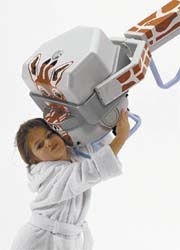
Equipment decorated with child-friendly motifs offers emotional rather than technical support in paediatric radiology. Some time ago, for its use at the Children’s Hospital Wilhelmsstift, Hamburg, Toshiba decorated an MRI scanner with the popular Janosch Design. The hospital’s mobile X-ray system Mobilett XP, made by Siemens, features a smiley giraffe. Masquerading as the giraffe’s neck, the machine’s large C-arm can be positioned for imaging in any projection.
Siemens’ syngoBlade, a complete imaging, matrix-based MRI software, ensures clear MR images even of agitated, fidgety little patients. Suitable for neurological and orthopaedic examinations, syngoBlade measures and corrects low-resolution images of each movement by continuous image-taking, to produce sharp images even of ‘busy’ little patients.
There are many ways of making radiology more suitable for children; however, nothing can replace a comforting word from the doctor and a small reward for bravery.
01.05.2007
More on the subject: