Neurology
By Dr Denis Ducreaux, Assistant Professor of Neuroradiology at CHU de Bicêtre, France
The value of diffusion-weighted and diffusion tensor imaging techniques.

DWI and DTI provide information on the mobility of water molecules in tissue. It is a widely accepted and utilised method for detecting acute ischaemic brain injury: highly mobile extracellular water shifts into the intracellular compartment generating ‘cytotoxic oedema’ during the early stage of arterial stroke. DTI using FT reconstructions may help visualise early wallerian degeneration in sub-acute and chronic stage of ischaemic stroke.
A major development has been achieved these past years in imaging spinal cord diseases. Several investigators have assessed the feasibility of performing spinal cord DTI studies. It is known that DTI sequences with computation of fractional anisotropy (FA) are more sensitive than spin echo T2 weighted images in detecting intrinsic abnormalities in acute or chronic spinal cord compression. In lesions that produce involvement of white matter fibres, it has also been reported that DTI with FT may potentially help to define abnormal areas that are undetected on routine T2-weighted imaging. FA and FT maps, derived from DTI computations, may help neurosurgeons to better delineate tumours and might contribute important information before tumour resection.
There are many clinical applications of DTI and FT. Recent ones focused on the spinal cord.
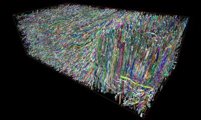
Normal spinal cord anatomy can be studied using FT and shows the main white matter tracts: posterior-lateral cortico-spinal, posterior lemniscal, and spinal-thalamic. On the fibre tracking 3D reconstructions, it is also possible to visualise bunches of fibres in the nerve roots.
In spinal cord tumours, DTI and FT may help to characterise and differentiate tumours (astrocytomas, ependymomas, haemangioblastoma, metastasis) and to delineate their margins.
In spinal cord acute and chronic compressions, conventional T2 weighted images may underestimate the effects of compressive lesions on the cord particularly when no hyperintense signal accompanies cord compression in the hyperacute period (the critical time period to treat these lesions). DTI can detect abnormal areas within a normal appearing spinal cord on T2-weighted imaging, and may also help to predict the patients’ outcome.
In myelitis, DTI is more sensitive than regular T2-weighted imaging in detecting spinal cord inflammatory lesions. Additionally, FT shows that inflammatory lesions spread the fibres of the spinal cord in areas that have abnormal T2 signal. This pattern is not seen in invasive tumours.
In metabolic disorders, such as MELAS (mitochondrial encephalopathy, lactic acidosis, and stroke like events) it is a disorder that affects both the brain and spinal cord. On the T2-weighted and FLAIR images multiple abnormal high intensity signal lesions may be seen in the mesencephalon, medulla oblongata, cerebellum and cervical spinal cord. FA values are decreased within the spinal cord of these patients, even when T2 abnormalities are not obvious.
DTI and FA are then more sensitive than other conventional MR imaging techniques to detect, characterise and map the extent of spinal cord lesions. In addition, FT offers the possibility of visualising the integrity of white matter tracts surrounding some lesions and this indirect information might help in formulating a differential diagnosis and in planning biopsies or resection.
17.11.2006


