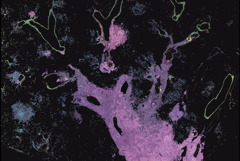
image credit: Dr. Eric Gardner, Varmus Lab
News • Histological transformation
How lung cancer can transform into another type
Lung tumors called adenocarcinomas sometimes respond to initially effective treatments by transforming into a much more aggressive small cell lung cancer (SCLC) that spreads rapidly and has few options for treatment.
Researchers at Weill Cornell Medicine have developed a mouse model that illuminates this problematic process, known as histological transformation. The findings, published in Science, advance the understanding of how mutated genes can trigger cancer evolution and suggest targets for more effective treatments.
The researchers discovered that during the transition from lung adenocarcinoma to small cell lung cancer (SCLC), the mutated cells appeared to undergo a change in cell identity through an intermediate, stem cell-like state, which facilitated the transformation. “It is very difficult to study this process in human patients. So my aim was to uncover the mechanism underlying the transformation of lung adenocarcinoma to small cell lung cancer in a mouse model,” said study lead Dr. Eric Gardner, a postdoctoral fellow in the laboratory of Dr. Harold Varmus, the Lewis Thomas University Professor of Medicine and a member of the Sandra and Edward Meyer Cancer Center at Weill Cornell Medicine. The complex mouse model took several years to develop and characterize but has allowed the researchers to crack this difficult problem.
This shows that an ‘oncogene’ in the wrong cell type doesn’t act like an oncogene anymore. So, it fundamentally changes how we think about oncogenes
Ashley Laughney
This study was in collaboration with Dr. Ashley Laughney, assistant professor of physiology and biophysics and a member of the Meyer Cancer Center at Weill Cornell Medicine and Ethan Earlie, a graduate student in the Laughney lab and part of the Tri-Institutional Computational Biology and Medicine program. “It is well known that cancer cells continue to evolve, especially to escape the pressure of effective treatments,” said Dr. Varmus. “This study shows how new technologies—including the detection of molecular features of single cancer cells, combined with computer-based analysis of the data—can portray dramatic, complex events in the evolution of lethal cancers, exposing new targets for therapeutic attack.”
SCLC most commonly occurs in heavy smokers, but this type of tumor also develops in a significant number of patients with lung adenocarcinomas, particularly after treatment with therapies that target a protein called Epidermal Growth Factor Receptor (EGFR), which promotes tumor growth. The new SCLC-type tumors are resistant to anti-EGFR therapy because their growth is fueled by a new cancer driver, high levels of Myc protein.
To unravel the interplay of these cancer pathways, the researchers engineered mice to develop a common form of lung adenocarcinoma, in which lung epithelial cells are driven by a mutated version of the EGFR gene. They then turned the adenocarcinoma tumors into SCLC-type tumors, which generally arise from neuroendocrine cells. They did this by shutting off EGFR in the presence of several other changes including losses of the tumor suppressor genes Rb1 and Trp53 as well as turning up the production of Myc, a known driver of SCLC.
Oncogenes, such as EGFR and Myc, are mutated forms of genes that normally control cell growth. They are known for their roles in driving the growth and spread of cancer. Tumor suppressor genes, on the other hand, normally inhibit cell proliferation and tumor development.
Surprisingly, this study showed that oncogenes act in a context-dependent manner. While most lung cells are resistant to becoming cancerous by Myc, neuroendocrine cells, are very sensitive to the oncogenic effects of Myc. Conversely, epithelial cells, which line the air sacs of the lungs and are the precursors to lung adenocarcinomas, grow excessively in response to mutated EGFR. “This shows that an ‘oncogene’ in the wrong cell type doesn’t act like an oncogene anymore,” Dr. Laughney said. “So, it fundamentally changes how we think about oncogenes.”
The researchers also discovered a stem cell-like intermediate that was neither adenocarcinoma nor SCLC. Cells in this transitional state became neuroendocrine in nature only when mutations in the tumor suppressor genes RB1 and TP53 were present. They observed that loss of another tumor suppressor called Pten accelerated this process. At that stage, oncogenic Myc could drive these intermediate stem-like cells to form SCLC-type tumors.
This study further supports efforts seeking therapeutics that target Myc proteins, which are implicated in many types of cancers. The researchers now plan to use their new mouse model to further explore the adenocarcinoma-SCLC transition, detailing, for example, how the immune system normally responds to this transition.
Source: Weill Cornell Medicine
12.02.2024


