Low dose CT wins the confidence of cardiologists
Patient referrals have doubled for CT angiography since 2004 as scanners cut radiation by 75%, yet produce ever-higher quality images, says John Brosky
He literally wrote the book on the subject, so when Jean-Louis Sablayrolles MD, states that low dose computed tomography angiography (CTa) represents a ‘great revolution in cardiac imaging,’ his words carry the weight of deep clinical experience.
and apical segments of the left ventricle
Appearing on the shelves of the medical publication specialist Springer during the 2010 European Society of Radiology (ESR) congress in Vienna, his 300-page book, Low Dose Coronary CTa in Ischemic Disease, provides practical guidelines on coronary artery imaging based on his work as the head of CT and MRI operations at the high-volume, 182-bed Centre Cardiologique du Nord (CCN) just outside of Paris.
Dr Sablayrolles told European Hospital the increasing acceptance of CTa by cardiologists can be seen in the growth curve of examinations performed at CCN, which quite simply have doubled from 1,500 per year in 2004, when the ‘revolution’ began, to more than 3,000 examinations in 2009.
CCN performs between 10 and 20 exams daily and, over the five-year period, has accumulated an experience of more than 11,000 exams on low-dose CT, which Dr. Sablayrolles drew upon in developing his recommendations for colleagues.
Radiation of patients has posed the greatest barrier to acceptance of CTa, he said, and it remains a key concern among cardiologists as a patient’s care path may require several examinations during the course of diagnosis, treatment and follow up.
Yet the upward curve of requested examinations can be linked directly to a decreasing curve that traces the reduction in X-ray energy required to produce high resolution cardiac images.
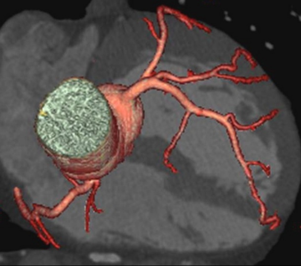
‘The introduction of 64 rows for the CT detector array made an enormous difference, providing excellent images of coronary arteries and plaque formations,’ said Dr Sablayrolles, ‘but there continued to be a great concern over the 16 millisievert (mSv) radiation dose that was required.’
The revolution arrived in a rapid succession of engineering advances that enable faster acquisition speeds, enhanced image processing, and an innovative technique for turning the radiation on-and-off during the cardiac cycle.
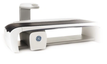
In November 2005, Dr Sablayrolles could report to cardiologists at the American Heart Association meeting that exam time had been reduce to five heart beats to produce what he called ‘remarkable cardiac images’ using a Lightspeed Volume CT scanner from GE Healthcare, which had been installed 11 months earlier. Significantly, he said, cutting acquisition time in half created an opportunity to examine a broader population of at-risk patients with rapid heart rates who previously were unable to tolerate the longer exam time.
More recently, a novel technique called ‘prospective triggered gating’ was added to the volume scanner, linking the pulses of radiation to the patient’s echocardiogram (ECG) so that the X-ray is turned on only during the optimum moment in a cardiac cycle, and then simply turned off when not acquiring useful images. With this innovation radiation exposure fell from 16 mSv to 8 mSv, he said, and cardiac volume CT exams became routine at CCN.
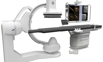
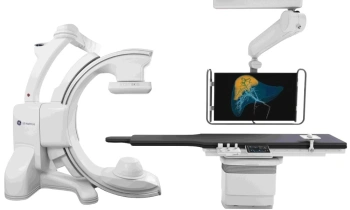
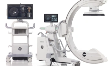
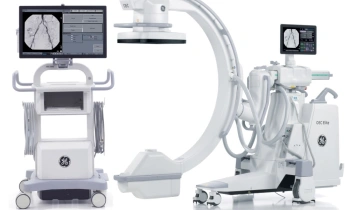
In late 2008, he said, CCN cut radiation exposure by more than half again, to between 2-3 mSv, with the installation of the Discovery CT750 HD, also from GE Healthcare, which, in the first five months of operation, was used for 2,500 exams.
With the excitement of a car enthusiast Dr Sablayrolles detailed the advanced electronics, enhanced ASIR algorithms for image processing and, most critically, the precision of the triggered gating process for image acquisition with radiation. ‘Already the great revolution in cardiac imaging was realised by dramatically lowering the dose and increasing image quality to a level that was never seen before at even the highest exposure rates,’ he said, ‘so that these new capabilities respond in double measure to the challenges.’
‘More and more as radiologists we are collaborating in therapeutic decision support with images of complex lesions, a topography of plaque formations, and an analysis of stent placement,’ he said. ‘We are helping to optimise procedures, whether for determining the choice of a stent, or for identifying extended areas of plaque that could provoke an incident during surgery.’
‘But nothing is perfect and there are still challenges to overcome,’ he pointed out. ‘You know we are never satisfied, and we need to go further.’
04.03.2010