Imaging of myocardial viability and perfusion with dual energy CT and dynamic DSCT
Authors: B. Ruzsics1 , T. Henzler2 , C. Fink2, S. O. Schoenberg2, Joseph U Schoepf1
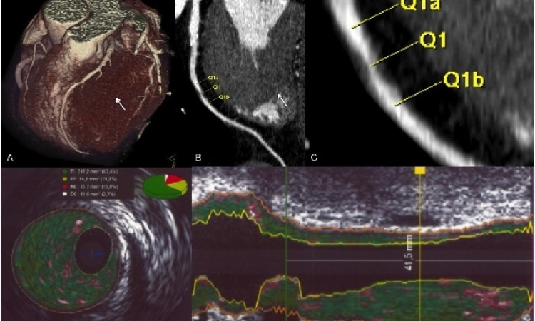
Although cardiac computed tomography (cCT) has shown very good diagnostic accuracy for the detection of coronary artery disease, the physiologic significance of many lesions can be uncertain. Furthermore, the presence of calcified atherosclerotic plaque reduces the ability to differentiate significant stenosis from non obstructive plaque.
Rapid evolution of multi detector-row computed tomography (MDCT) technology continues to enhance the role of this technique in the work-up of coronary artery disease. Recently, MDCT has been proposed as a stand-alone modality for integrative imaging of coronary heart disease, i.e. the comprehensive assessment of cardiac anatomy, function, perfusion, and viability. Single-energy and dual-energy CT techniques based on dual-source CT (DSCT) technology have been proposed for this purpose.
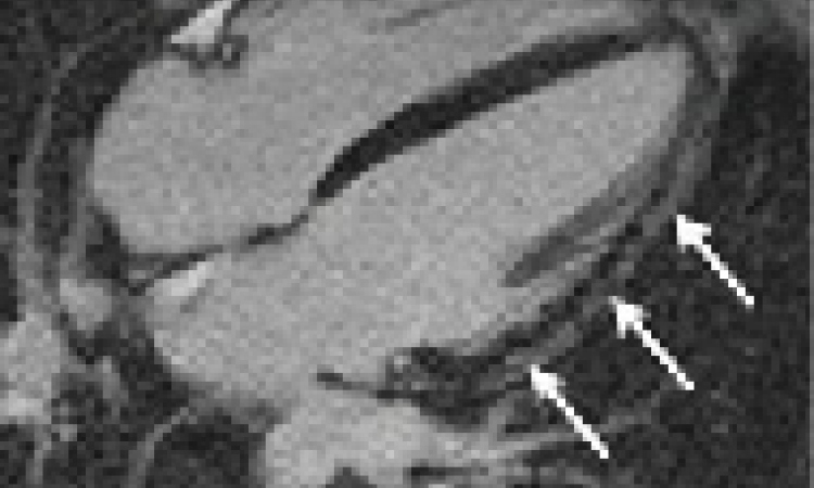
DECT has the potential to evaluate changes in the status of the myocardial blood supply in addition to the analysis of coronary artery morphology. It has to be mentioned that current DECT approaches are based on static non time resolved coronary CT angiographic scans. DECT exploits the fact that tissues in the human body and the intravascular iodine-based contrast media have unique spectral characteristics when penetrated by different x-ray energy levels. This property enables the mapping of the iodine (and thus blood) distribution within the myocardium demonstrating the hemodynamic significance of non-invasively detected coronary artery disease with a single contrast-enhanced exam. In a recently published study by our group, ECG-gated DECT of the heart showed 92% accuracy for detection of >50% stenosis and an accuracy of 94% for the detection of myocardial ischemia compared to single photon emission computed tomography (SPECT).
A drawback of DECT acquisition of the heart is that the temporal resolution of DSCT decreases from 83 to 165 ms, because the 2 tubes that enable quarter-scan reconstruction during routine, DSCT coronary angiography are used to generate different energy spectra in DE mode. However, the temporal resolution of DECT is identical to that of conventional single-source 64-slice CT scanners. In order to preserve the high temporal of DSCT we have recently evaluated the feasibility of a novel hybrid reconstruction algorithm for DECT which combines both, quarter and half-rotation reconstruction with high- and low-pass filtering techniques. The hybrid reconstruction algorithm used in this study had a 91.4% sensitivity, 94.7% specificity, 82.1% positive (PPV), and 97.7% negative predictive value (NPV) for detecting significant stenosis versus 85.7%, 93.2%, 76.9%, and 96.1% with a standard DECT reconstruction algorithm.
Adenosine-stress dynamic myocardial volume perfusion with second generation DSCT
Recently, second generation DSCT was introduced which compromises two 128-section detectors that provide greater coverage compared with first generation DSCT. This system provides the ability of performing dynamic perfusion imaging by means of a dedicated ‘shuttle’ mode that captures the passage of a contrast medium bolus through the myocardium.
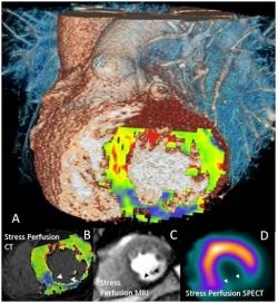
In the ‘shuttle’ mode of second generation DSCT (SOMATOM Definition Flash, Siemens Healthcare, Forchheim, Germany) the table shuttles back and forth during image acquisition between two adjacent anatomic positions with 300 mm/s acceleration. This acquisition mode can also be performed in an ECG-triggered fashion. Given a detector width of 38 mm, and a 10% overlap between both acquisition ranges, the anatomic coverage of this imaging technique is 73 mm. In a recently published study we applied this acquisition technique for the purpose of myocardial perfusion assessment during contrast medium infusion and under pharmacological stress with adenosine as a component of a comprehensive CT based protocol for integrative imaging of cardiac structure, function, perfusion, and viability. In this study we found a strong correlation between stress first-pass myocardial perfusion and delayed enhancement imaging with CT and the myocardial perfusion assessed on stress MRI and delayed enhancement MRI.
In conclusion, both static DECT ‘perfusion’ and dynamic myocardial volume perfusion imaging techniques are novel promising tools of cCT angiography which allow estimating the significance of atherosclerotic lesions in a single examination. DECT has the advantage of a lower radiation dose and a single contrast material administration whereas dynamic DSCT volume perfusion allows real time resolved rest and stress imaging of myocardial perfusion.
Additionally, we would like to invite you to the second transatlantic ACSI meeting, on 25-26 June 2010 in Mannheim, Germany.
This transatlantic symposium is held annually, alternating between a transatlantic partner and Mannheim, Germany. Our partner this year will be the Clínica de Diagnóstico por Imagem (CDPI), Rio de Janeiro in conjunction with the Department of Diagnostic Radiology of the University of Rio de Janeiro.
Four topics (MRI, CT, PET and imaging economics) will be presented in a two day programme by international experts in clinical imaging as well as technical and economic developments.
We are pleased to offer you a very attractive and innovative programme and look forward to welcoming you in Mannheim this June.
Further information: www.mr-pet-ct.com
1. Department of Radiology and Radiological Science, Medical University of South Carolina,
Charleston, South Carolina
2. Department of Clinical Radiology and Nuclear Medicine, Mannheim University Medical Centre, Mannheim Medical Faculty, Heidelberg University, Germany
25.05.2010