Hot topic cardiovascular imaging
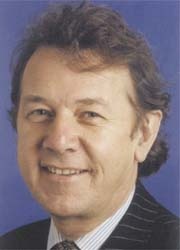
Every summer the European Society of Cardiology (ESC) holds Europe's biggest annual meeting of specialists in cardiovascular medicine, inviting and drawing in top international medical professionals. Karoline Laarmann asked Professor Kim Fox, President of the European Society of Cardiology and Consultant Cardiologist at the Royal Brompton Hospital, and professor of clinical cardiology at Imperial College, London, UK, about the most relevant issues and highlights planned for this year's programme.

HPS, and ASCOT etc, which have revolutionised the treatment of this condition. He also chairs the task force producing the European Angina Guidelines as well as the committee responsible for the audit for the management of these patients in Europe.
Prof Fox has published over 400 papers in leading scientific journals, lectured to national societies worldwide, and has given the Finlayson Lecture to the Royal College of Surgeons in Glasgow, the St Cyres Lecture to the British Cardiac Society and the Andrea Cissalpino Lecture to the Italian Society of Cardiology.
For his services to cardiology he is an elected honorary member of the French Cardiac Society and the Japanese Circulation Society.
Last year’s key focus was on heart failure. What will be this year’s hot topic?
KF: ‘This year we decided to go for cardiovascular imaging because any cardiovascular condition that will be discussed during our congress always involves imaging in some form. Imaging is the cornerstone not only for diagnosing a condition but also to assess prognosis and tailoring individual treatment, too. By imaging we are talking about procedures from the simplest form such as the electrocardiogram to the most complicated non-invasive imaging techniques – MRI, nuclear imaging, echocardiography and, more recently, cardiac CT scanning, and of course not forgetting to mention all the invasive techniques: cardio-angiography and cardiac catherisation.’
Which of the current imaging modalities is making the strongest progress?
‘The gold standard is multimodality imaging, because although a single tool may be sufficient, patient evaluation is often best when used in conjunction with other techniques. Every imaging technique has its individual strengths and weaknesses; they all give a different piece of information, so it depends on what you intend to measure.
Therefore one of the hot ESC topics will be to discuss which are the right imaging tools to use for a special problem. Of course we have considerable hopes in molecular imaging. It may be the tool to identify different forms of heart muscle diseases that may respond to different therapies and, in particular, may respond to stem cells. If it were to be shown that stem cells were to be beneficial, then to deliver the stem cells will require sophisticated imaging. The second hope we nourish in molecular imaging is to identify vulnerable plaques. We’ve just started to get some imaging information by using intravascular ultrasound of the coronary artery.’
What form does work sharing take between cardiology and radiology imaging?
Nowadays the management of a patient is multidisciplinary, to achieve the best quality of healthcare. As a cardiologist I think that radiology is extremely important in particular cases where physics is involved – radiation in particular. Radiologists need to be involved in ensuring that proper safety is taken into account. However, when it comes to actually deciding which imaging tool should be used for a patient, the cardiologist must be the person to make the decision. The most controversial issue here is: Who should execute the procedure? The demarcation lines for that are changing currently and they differ from country to country. In the UK, for example, the radiologist will lead on cardiac CT, most of the others by cardiologists but with special expertise in imaging. I assume that coronary CT will probably remain the area of radiologists just now.
What will be the biggest challenges for cardiology in the future?
‘Heart transplantation is a fantastic technique but we clearly need to think about some form of alternative technique, which may be stem cells or a form of artificial heart. Better still, of course, would be better implementation of prevention. We probably could deal with most of the cases of heart failure if prevention would be better, which brings me to obesity, which causes an increasing number of heart diseases. Last year, at the ESC opening ceremony, I emphasised my dislike of smoking. Now
I see child obesity as a major issue to take care of. But it is much tougher to handle because you cannot legislate what people eat. The trouble is that fast food is much cheaper than a healthy diet. It will take huge educational programmes to accomplish a change of thinking.
‘Finally, it would be very helpful to identify the 20–30% of patients benefiting from treatment with drugs, because this would lead to enormous cost savings and allow access to medicinal treatment for people who truly need it. What we are doing now is treating the whole population with a drug or procedure from which only a few profit. But the most important thing to do in cardiology is prevent ischaemic heart disease. We have to identify the people who show an aggravated risk to optimise our strategies of prevention and treatment based on the individual.’
01.09.2008


