Hot on the heels of cardiac catheterisation
The multicentre international trial CorE-64 trial has demonstrated that CT is increasingly important in the non-invasive diagnosis of obstructive coronary artery disease.

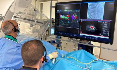
For the first time, radiologists have tested the reliability of cardiac CT results compared to cardiac catheterisation in this multicentre international trial (carried out by the Charité University Clinic in Berlin and Johns Hopkins University, USA, among others). The result: Non-invasive CT facilitates the safe detection of vasoconstriction; however, for a precise assessment of the severity of obstructions, the catheter was still superior to CT enhanced imaging. ‘The result makes us feel confident. The study shows that CT is hot on the heels of angiography,’ said lecturer Dr Marc Dewey (above), of the Institute of Radiology, Charité Mitte Campus, Berlin, who led the study in Germany. ‘Catheterisation is not without its risk. Deposits removed from the vascular walls can lead to constrictions in other parts of the organ, or the vascular walls can actually tear,’ he pointed out.
Unsurprisingly, about 75% of patients stated they would prefer fast and painless CT for future examinations. According to the radiologist only a third of catheterisations are actually combined with therapy, i.e. the vessels are only widened in about a third of cases. In all other cases, catheterisation is only used for clarification.
Seeking a low-impact diagnostic procedure
For some time, radiologists have been looking for a way to assess reliably the condition of coronary arteries whilst avoiding the complications implicated in doing this via angiography. CT has proved to be the procedure with the biggest potential to take over from catheterisation, as the international team carrying out the CorE-64 study discovered.
Some 400 patients with diagnosed or suspected coronary heart disease underwent a two-fold examination (approved by the Federal Office for Radiation Protection), employing catheterisation and CT. ‘We were able to detect stenosis requiring treatment using CT with the same precision as with the cardiac catheter, which means non-invasive CT examination that is free of complications is equally capable of identification as catheterisation,’ Dr Dewey concluded.
‘64-slice CT is currently the most promising procedure for non-invasive coronary angiography. However, in a direct, multi-centre comparison with conventional coronary angiography limitations are also evident. The sensitivity of 85% and the negative predictive value of 83% are lower than in previous single-centre studies,’ he explained. ‘This means that, with large-scale use of 64-slice CT for coronary angiography, the realistic results for diagnostic precision are lower than in previous examinations (with about a 95% sensitivity) which were not multi-centre. Nevertheless, and this is clearly demonstrated by our first international, multi-centre study, the diagnostic value of CT is higher than with all other non-invasive procedures to detect coronary artery stenosis. Moreover, CT was on a par with conventional coronary angiography in the prediction of the necessity of coronary revascularisation.’
In terms of assessing for which risk score cardiac catheterisation and CT are appropriate Dr Dewey added: ‘Non-invasive cardiac CT could be a sensible “filter” prior to cardiac catheterisation for patients with low to medium probability (20-60% pre-test probability) of coronary artery stenosis. For example, these are patients with asymptomatic complaints or existing, conflicting results of other examinations. Patients with typical, symptomatic problems should definitely still be examined via cardiac catheterisation and, if necessary, revascularisation. On the other hand, it has been shown that there is no improvement in clinical, long-term results in asymptomatic patients, which means that CT does not seem to be a sensible approach for these patients. As Rita F Redberg writes in her article about our study in the New England Journal of Medicine, there is a need for more randomised – and ideally publicly sponsored – studies to further analyse the benefits of low-impact, non-invasive CT prior to the large-scale use of cardiac CT.
* The CorE-64 results were first published in the New England Journal of Medicine (2008; 359: 2324-36).
20.12.2008


