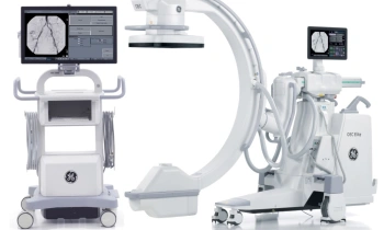
GE high-definition CT technology
High definition CT (HDCT) technology developed by GE Healthcare promises to revolutionise image acquisition for CT scanning.
Dr Dennis Foley, Director of Imaging at Milwaukee’s Froedtert Lutheran Medical Center and the Medical College of Wisconsin was the first to utilise a number of HDCT technologies. Here, for European Hospital readers, Dr Foley reports his experiences.
HDCT technologies are a portfolio of developments that improve the hardware, software and electronics of the system. From the hardware, GE designed a new scintillator, which should perform dual energy with almost simultaneous acquisition of the projection data at different beam energies. Due to rapid beam energy switching and electronic readout within 8ms, almost identical projection angle data at the two different beam energies can be obtained. This is much faster than a system that is fundamentally mechanical in approach and has two X-ray tubes inside the gantry, which results in a 83ms delay in obtaining the same projection angle at different beam energies.
Potentially, the new scintillator can enable subtraction imaging of coronary artery calcification – a major breakthrough. Today, the challenges in coronary CT are improved temporal and spatial resolution, in addition to the removal of artifacts. Artifacts relate largely to coronary calcification. When performing selective coronary arteriography, subtraction imaging is an integral part of the cardiologists process. If CT can emulate selective coronary catheterisation by removing calcification, this would be a major advance.
Prospective gating for cardiac CT has already been implemented for GE Healthcare’s LightSpeed system. This technique which is fundamentally a step-and-shoot technique, and is applied in patients with regular heart rate, produces images equivalent to those obtained with retrospective gating, but with significant reduction in radiation dose. Radiation dose is reduced as data is obtained only at a predefined point in each R-R interval, compared with retrospective gating in which the X-ray tube is on continuously, throughout the cardiac cycle. Prospective gating reduces radiation dose to approximately one half of that obtained with retrospective gating, even with EKG gated tube current modulation. Prospective gating requires a system with adequate beam width and appropriate software.
A new approach to CT image formation is iterative reconstruction. This is a relatively software intensive approach in which images that are smooth, and have good sharp anatomic outlines, are obtained at about one half the radiation dose utilised for conventional CT scanning.
Volume dual energy techniques for cardiac and non-cardiac imaging and iterative reconstruction remain in clinical developmental phases and implementation depends on software engineering and initial clinical applications. However, I am optimistic that these technologies will come into clinical practice within the next 12 months.
09.03.2008