Gadolinium-containing contrast materials
"Why all the fuss in 2007?" asks Peter Gross MD, Professor of Medicine and Nephrology at the C G Carus University Hospital in Dresden
Medical imaging is a first order boom area.

In many instances iodine-containing contrast materials (I-CM) are highly efficient in improving the diagnostic power of imaging even further. An I-CM is disposed of by the kidneys. Due to the renal concentrating mechanism, this results in renal intratubular levels of I-CM that are multi-fold (up to 100-fold) higher than their concomitant plasma levels. Healthy kidneys take this challenge in their stride. However, damaged or impaired kidneys (chronic renal failure; diabetic nephropathy; proteinuria; dehydration; presence of hypertension or congestive cardiac failure; old age) will often react differently and go into acute renal failure (ARF is also termed acute kidney injury) temporarily. While the incidence of ARF after I-CM in normal kidneys approaches zero, the incidence may be 3-20% in patients with damaged or impaired kidneys, depending on the number of attendant risk factors. A few prophylactic measures against this ARF have been established: appropriate plasma volume expansion with 0.45 or 0.9% NaCl before I-CM and use of iso-osmolar, non-ionic I-CM (e.g. iodixanol) for the respective X-ray studies or CTs. Nonetheless, the absolute numbers of I-CM induced ARF apparently have not fallen. This appears to be explained by the ever increasing ‘load’ of risk factors in today’s patient population (increasingly old, the increase in diabetes mellitus and more frequent chronic renal failure, etc) offsetting progress made in prophylaxis. Hence the intense search for alternative techniques and CMs
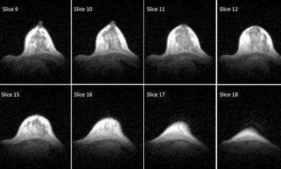
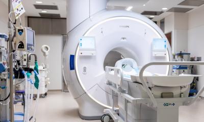
In this context magnetic resonance imaging (MRI) and Angio-MRI, both using Gadolinium containing CM (Gd-CM), have received considerable interest. It became standard procedure to refer patients at significant risk for I-CM induced ARF (after X-ray studies) to Gd-CM involving MRI whenever possible. This appeared justified by research demonstrating a marginal or even non-existing incidence of ARF after Gd-CM in patients with risk factors – as long as a maximal dose of Gd-CM of 0.3 mmol/kg BW was heeded. (At this dose, imaging contrast in MRI is sufficiently excellent).
The ‘blind confidence’ in the non-toxic nature of Gd-CM in patients with risk factors went further. Radiologists proceeded to use Gd-CM for X-ray studies such as angiography and CT. However, Gd-CM had been developed for contrast under conditions of magnetism not radiation (X-ray). Hence technical limitations forced a 2-6-fold increase of the dose of Gd-CM when used ‘off-label’ in X-ray studies; otherwise adequate imaging would be unattainable. Therefore, it was frequently possible that the upper limit of recommended Gd-CM dose of 0.3 mmol/kg BW would be transgressed. Nonetheless, the use of Gd-CM for X-ray, and often at these high doses, was considered acceptable.
Given these circumstances it is not surprising that a backlash occurred. On the one hand several recent reports suggested that Gd-CM may also occasionally cause ARF in patients with risk factors. It is true that these kinds of ARF are mostly reversible and short lasting; however, they are costly, burdensome and prolong hospitalisation. Above all else, they are in a way iatrogenic. The aura of Gd-CM now has a shadow.
On the other hand, an obviously pernicious new syndrome has been reported, with increasing frequency, mostly in the last two years: Nephrogenic systemic fibrosis (NSF). Its occurrence appears to be limited to only a few – reportedly 2–3 % – of those patients that received Gd-CM while having an estimated glomerular filtration rate (eGFR) <30 ml/min (some say <60-30 ml/min) i.e. in patients with a significant risk factor burden. Conversely, NSF has been almost totally absent from chronic renal failure patients who had never had any exposure to Gd-CM. Those affected by NSF suffer what is vaguely reminiscent of scleroderma. There will be increased and hard connective tissue primarily in the extremities. In some patients the condition may cause an inability to use hands or changes involving the feet may confine them to a wheelchair; in others NSF may progress to involve internal organs, potentially leading to death from pulmonary or cardiac failure.
At this time a little over 250 cases of NSF have been described worldwide. The causation of NSF by Gd-CM has not been proven rigorously and some believe that it is multifactorial in nature. However, the context of NSF and Gadolinium is highly suggestive and hence most physicians are convinced that the relation will turn out to be real. The pathogenesis has remained largely unclear. There is no known therapy that works reproducibly. In terms of potentially triggering NSF, it is uncertain whether or not some of the 6 Gd-CM presently on the market are ‘safer than others’. Most observers believe it is rather a class effect of all Gd-CM that is involved in NSF.
German health authorities (BfArM) have embargoed some Gd-CM, meanwhile (Omniscan, Magnevist) when there is an eGFR <30 ml/min. (Omniscan and Magnevist are the 2 Gd-CM with the biggest world market share at this time). In the same context, the BfArM warns against the use of the other Gd-CM. Health Authorities in many other countries have been quick to install comparable measures in the past.
In the future more scientific data will be necessary; we must learn the pathogenesis and we have to find efficient approaches to prophylaxis and therapy. Imaging and contrast enhancement are now essential to physicians and highly advantageous to patients, so their further progressive development must not be slowed by serious adverse events, such as those described here. Towards those ends a registry for cases of NSF has just been established in Germany by the Nephrologische Gesellschaft. In the USA a registry of this kind has been in existence for a little longer (Yale University Medical Centre).
30.10.2007


