Image source: Griffith University
News • Non-invasive testing
Early diagnosis of lung cancer from breath samples
The human breath could help identify cancerous lung nodules negating the need for invasive testing.
New Griffith University research, funded by Tour de Cure, analysed exhaled breath to help doctors distinguish between cancerous and non-cancerous lung nodules, supporting earlier lung cancer diagnosis. Approximately 2.5 million new cases of lung cancer are diagnosed each year.
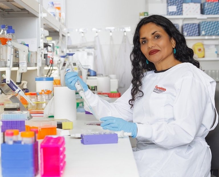
Professor Chamindie Punyadeera from Griffith’s Institute for Biomedicine and Glycomics said the study evaluated two complementary technologies, with both showing promise. “We studied an electronic nose, or eNose, which identifies patterns in breath chemicals,” Professor Punyadeera said. “The second technology was gas chromatography-mass spectrometry which identified specific volatile organic compounds in breath. Both breath-based approaches showed substantially better discrimination between malignant and benign nodules than models using standard clinical information alone.”
While a simple breath test is showing promise in identifying which nodules are most likely to be malignant, it is not intended to replace biopsies, but rather to help clinicians decide who needs invasive intervention most urgently
Xi Zhang
The research could replace the need for invasive testing which currently entailed a CT-guided lung biopsy to obtain tissue for a diagnosis. Other invasive techniques encompassed a bronchoscopy to sample suspicious nodules, or in some cases, surgery to remove or investigate nodules that may be cancerous.
Sometimes patients underwent invasive techniques to later find out 95% of pulmonary nodules identified on low-dose CT scans were ultimately found to be non-cancerous.
Co-author Dr Xi Zhang said: “Biopsying lung nodules can be technically challenging, particularly when lesions are small or difficult to access, and can cause complications such as pneumothorax or collapsed lung. While a simple breath test is showing promise in identifying which nodules are most likely to be malignant, it is not intended to replace biopsies, but rather to help clinicians decide who needs invasive intervention most urgently. It is hoped it could also potentially reduce unnecessary procedures for patients with benign nodules by including the non-invasive test into routine clinical care.”
Recommended article

Article • Breath analysis to aid diagnoses
Breathomics: far more than hot air
In diagnostics, it sometimes makes sense to follow your nose. During the Labmed Forum at Medica, Dr Beniam Ghebremedhin and Dr Simona Cristescu discussed the diagnostic potential of breathomics – the analysis of a patient’s exhaled air for disease indicators.
This research was conducted by PhD student Waqar Ahmed Afridi. Professor Punyadeera and Dr Zhang worked in collaboration with Adjunct Associate Professor David Fielding, a staff thoracic physician at the Royal Brisbane and Women’s Hospital, who provided clinical guidance and leadership throughout the research, was critical to the paper’s translation relevance. This research was undertaken in collaboration with Dr David Paas from Queensland Health.
Source: Griffith University
29.09.2026