Demand for PET/CT systems soars
Dr Thomas Beyer: `PET/CT is a non-invasive imaging modality that captures anatomical and metabolic data in a single scan´
No stand-alone PET units have been produced and sold for about two years, which means those who had already purchased a PET will now want a PET/CT*, according to Dr Thomas Beyer, International Manager of Clinical Sciences Nuclear Medicine & Pre-Clinical Imaging at Philips Medical Systems. ‘Physicians are demanding PET/CT systems because they are impressed by the quality of this imaging modality. However, in Europe, recognition of and reimbursement for PET/CT in Germany and Austria lag behind, even though, worldwide, it is now an established, standard procedure in oncology.’
In this field, it is used in the diagnosis of lung, oesophagus and colorectal carcinoma, lymphoma and melanoma as well as ENT and gynaecological tumours. However, PET/CT is also increasingly used to clarify cardiological and neurological issues. In terms of percentages, usage is 90 % oncology, 7 % cardiology and 3% neurology.
The future
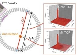
Very soon PET/CT systems will provide the full range of examination protocols already possible with stand-alone CTs and PETs — for example, triggering the scan according to the heartbeat, which reduces movement artefacts. We also anticipate protocols for radiation therapy planning. More than likely, PET/CT scans will become faster, which means very soon whole-body scans will be performed routinely and in less than ten minutes, thanks to technological developments, which include more sophisticated time difference measurements, for example the time of flight functionality.
*Positron emission tomography in combination with computed tomography
30.10.2007