CT: Optimising dosage
By Dr Jörg Blobel
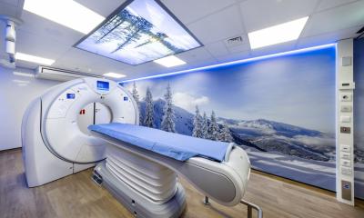
AquilionONE is the first CT scanner capable of imaging whole organ regions up to a width of 16 cm in one rotation and within a split second. Based on the raw volume data, rapid dynamic processes within an entire organ (e. g. heart, pancreas, kidney or brain) may be diagnosed with a time interval of 50 ms, i.e. with a rate of 20 volumes per second.

With their smallest effective width of 0.5 mm the detector elements ensure best possible spatial resolution. Image reconstruction of 640 slices, coupled with special mathematical interpolation, provides a geometric resolution of 0.4 mm image voxels in all directions. Unlike in flying-focus spot technology, the signal strengths are not halved and spreadover both slice series. Consequently, this halved signal strength per slice does not have to be compensated by increasing the exposure.
Heart rate adapted temporal resolution between 50 ms and 175 ms allows scanning of the entire organ within one heart beat. For rather high heart rates the temporal resolution of 50 ms is almost half that of a dual source CT scanner and the heart is better locked into position during motion. Compared with a Helical CT unit where the heart volume is captured by individual overlapping rotations the volume scan reduces the effective dose of the normal patient to 1.5-6 mSv – a reduction of 60-80%. Also the disadvantages of the step-and-shoot mode of multislice CT with a scanned field width of 20-40 mm, i.e. the common stepping artifacts at the volume borders and dose doubling along the volume overlap, are overcome. Data capture with Aquilion ONE requires just one heart beat. The modality is robust and offers increased potential to study arrhythmia patients. Calcified and non-calcified plaques, the latter frequently resulting in myocardial infarction, can be visualized down to a diameter of less than 1 mm.
This outstanding low-contrast resolution of the Aquilion ONE allows low radiation energy levels at 80 kV, e.g. for diagnostic organ perfusion scans. For the first time perfusion of the entire cerebral volume can be captured simultaneously. The 15-20 volume data sets generated during one minute of the study are acquired with a regular effective patient dose of just 4-6 mSv. Volumetric acquisition permits anatomically accurate fusion of the CT angiography and perfusion volumes (Fig1). The innovative range of CT studies opens up new perspectives for functional diagnostic workup, e.g. in joint movement, peristalsis, dynamic blood flow analysis and perfusion of numerous organs.
In diagnostic pediatric scans the radiation field and the number of detector element rows is collimated to the size of the organ. This rapid examination avoids the need for breathhold in infants and small children otherwise required by the longer scanning times. If an emergency thoracic CT study is needed in an infant the effective dose at a tube voltage of 80 kV is minimal at 0.16 mSv. Large areas of the body in combination with cardiac CT can be captured by individual volume scans much more rapidly than with helical CT and are subsequently “stiched” to one patient volume. Complex studies of the heart, lungs and the head may be combined with supplementary CT angiography and – based on the study plan – are optimized by automatic system selection of the scan parameters to reduce radiation exposure. Before the start of the study the expected patient radiation exposure is displayed and can be controlled.
Conventional helical CT mode with the selection of 16, 32 and 64 detector rows is also available and the volume scan mode is enhanced by numerous new dynamic function options. These innovations of the Aquilion ONE will alter the patient workflow between the diagnostic imaging modalities in radiology.
By Dr Jörg Blobel, Ph.D., Chief Clinical Science, CT Systems Division, Toshiba Medical Systems Corporation, and Jürgen Mews, of the CT Systems Division at Toshiba Medical Systems in Neuss, Germany
28.10.2008


