Cardiovascular MRI
Scientific studies show improved diagnoses and outcomes for acute myocardial infarction patients
The role of magnetic resonance imaging (MRI) to assess the effect of therapy in patients with acute myocardial infarction was demonstrated in a series of papers during the 12th Annual Scientific Sessions of the Society for Cardiovascular Magnetic Resonance (SCMR), held in Orlando, Fla. USA (29 Jan - 1 Feb).
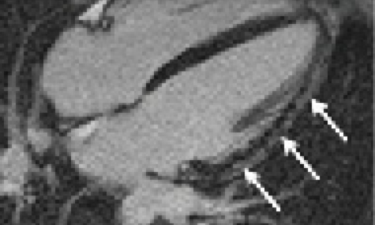
After therapy, cardiovascular MRI enables the measurement of the size a myocardial infarction would have been if no therapy had been performed (Salvage-imaging). Using this, physicians can assess the effect of their intervention and thus perform studies to optimise the benefit to patients. Prior to the arrival of this new technique this insight could only be obtained by comparing large groups of patients who had undergone different treatments to average the differences between the groups. The new method allows correction of these differences, promising quicker progress towards individually optimised therapy. Using it, said Holger Thiele MD (University of Leipzig, Germany) ‘…we have been able to show in a relatively small trial of 220 patients, that therapy in patients after myocardial infarction can be optimised adding anti-oxidative agents to standard therapy. We had known this from animal data before and were now able to demonstrate this effect in patients using MRI as an endpoint.’
MRI has already been established as an endpoint for cardiac function, size and measuring the size of irreversible myocardial damage after myocardial infarction. It is now expanding to a further endpoint for myocardial salvage in patients with acute myocardial infarction.
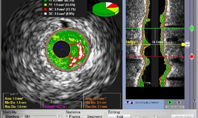
Perfusion imaging
The technique allows measurement of blood flow to the heart muscle with an unprecedented spatial resolution and without any ionising radiation. Multicentre data has already suggested a superiority of this technique in comparison to methods now used on a routine basis. New data, presented during the meeting, further expands its role, suggesting that new techniques with faster imaging will further improve spatial resolution and enhance diagnostic accuracy.
MR perfusion imaging, for example, may also be used in patients after bypass surgery because of its ability to discriminate areas with scar from the complex flow patterns seen after surgery. Similarly, the high spatial resolution allows detection of remaining areas of reduced blood flow after treatment. These new areas for use for MR perfusion imaging expand the indications of this relatively young technique and lead to a reconsideration of the application of MRI mainly as a second line technique.
Christoph Klein MD (German Heart Institute Berlin) pointed out: ’Given the amount of information we get from a single examination, ranging from the size and function of the heart, through scar tissue after myocardial infarction, to an excellent visualisation of myocardial blood flow, we should use MRI as the premier imaging technique in a large group of patients.’
Further research is guided towards quantitative assessment of myocardial blood flow, to allow doctors not only to discriminate between normal and abnormal blood flow but also to measure the effect of therapy and to individually guide patient management.
Registry reveals diagnostic value of MR scans
Data from a German registry on the utilisation of cardiovascular MRI in clinical practice, which involved almost 10,000 patients, demonstrated that this technique produced a final diagnosis in more than 70% of patients. 16% of the scanned patients had a different diagnosis than what was expected before their scans. The numbers are even more convincing when the complexity of the patients is considered.
Most of the patients had already received several other diagnostic tests before the cardiovascular MR examination. These numbers further underline the value of cardiovascular MR in clinical cardiology practice. In addition, the use of cardiovascular magnetic resonance as a first line technique, rather than being a late diagnostic procedure.
The registry is to be extended to an international level with participation of over 50 major diagnostic centres.
* This article is based on a sessions report supplied by SCMR
01.03.2009