Digital Pathology
CAD in pathology
Computer-aided diagnosis could soon play a greater role in digital pathology. Dr Jeroen van der Laak, an Assistant Professor in Digital Pathology at Radboud University, believes a breakthrough that would increase the speed and accuracy of diagnoses and prognoses is closer than many observers think
Report: Mark Nicholls
Van der Laak has been leading studies in the use of computer aided diagnosis (CAD) in pathology and will outline the ‘progress on the introduction of these promising techniques’ at the Digital Pathology Conference in London at the end of this year (3-4 December).
In his presentation ‘CAD in Pathology: Closer than You Think’ he will argue that, while the introduction of digital pathology in primary diagnostics is still hampered by high costs, CAD techniques will be the tipping point, leading to large scale introduction of digital scanning of histopathological slides.
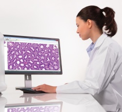
The introduction of CAD in pathology will make digitisation of tissue sections a worthwhile step, he believes. ‘As it is now, many pathologists doubt this transition to “digital pathology” because the required investment is large, and benefits are not fully clear,’ he said. ‘If we can really take some of the workload off a pathologist - by pre-screening sections for certain tedious tasks, for example - the business case will become much more favourable.’
Significant steps are achieved
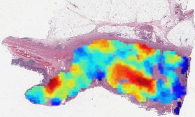
Research in his department – undertaken mainly using a Pannoramic P250 scanner from Budapest-based firm 3DHistech with all pattern recognition software developed by the group – has achieved significant steps in the last couple of years in developing advanced pattern recognition algorithms that can work on entire ‘whole slide images’ (WSI), in contrast to much research still being conducted on small image regions.
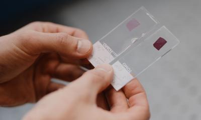
‘We also aim to use standard H&E staining whenever possible. This means that the software we develop will work in any lab, without special adjustments, and with hardly any user interaction.’
However, although the introduction of CAD in pathology is much closer than many people realise, he acknowledges that the availability of algorithms is only a new beginning and further research is needed to bring the algorithms into the pathologists’ workplace. ‘If we succeed in seamless integration in to daily pathology routine,’ he said, ‘and can prove that the software really aids the diagnostic process, large scale introduction may happen.’
Such a step would see pathologists become more efficient in certain routine tasks, and it could improve the accuracy and reproducibility of diagnosis, he suggested.
‘Future applications may add valuable information from tissue sections that is currently not available. Advanced pattern recognition may enable extraction of strongly prognostic, sub-visual information,’ van der Laak pointed out. ‘CAD in pathology has the promise to radically change the way we examine tissue sections.
‘We are at a stage in which we have availability of pattern recognition techniques more powerful than ever before. What is needed now is availability of material from large cohorts of patients with treatment and follow-up data.
‘Trained pathologists are also needed to help identify regions of interest and to study how we can best include our algorithms in the diagnostic workflow.
‘These requirements are challenging because pathologists frequently suffer high workload, and well-defined cohorts are often hard to acquire.’ A next step is for his group to study the benefit of developed algorithms in a routine diagnostic setting.
Yet, if implemented, CAD in pathology could have benefits for pathologists in making routine tasks easier. Advanced quantification of histopathological patterns will add valuable information to the diagnostic process, he said.
Patients will benefit because the diagnostic process may be more accurate and personalised, and surgeons and oncologists may also benefit from increased speed and accuracy of diagnostics and prognostics.
PROFILE:
Dr Jeroen van de Laak is Assistant Professor in Digital Pathology at the Department of Pathology at Radboud University Nijmegen Medical Centre, The Netherlands, where he leads a research group on CAD in pathology. Currently numbering eight, the group is set to expand in 2016. Covering diverse disciplines – computer science, medicine, lab technology, the group is embedded in the pathology department, and included in the radiology department’s diagnostic image analysis group (DIAG) at Radboud University Medical Centre.
04.09.2015