News • Prognosis and diagnosis
Deep insight into the heart
By no means are only elderly people at risk from heart diseases. Physically active individuals can also be affected, for example if a seemingly harmless flu bug spreads to the heart muscle. Should this remain undetected and if, for example, a builder continues with his strenuous job or an athlete carries on training, this can lead to chronic inflammation and in the worst case even to sudden death.
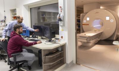
The latest issue of the “Forschung Frankfurt” journal describes how modern non-invasive examinations using state-of-the-art imaging technology can reduce such risks. Professor Eike Nagel and his 12 coworkers at the Institute for Experimental and Translational Cardio Vascular Imaging of Goethe University Frankfurt are developing better ways to predict and diagnose heart diseases.
In recent years, the researchers have taken the lead in the development of a procedure that is still very new in heart scans. Nagel explains the advantages: “With the help of magnetic resonance imaging, we can look right inside the heart muscle.” Blood flow to the heart muscle is visualized and shows whether there are any constrictions of the arteries supplying the heart. Experts can also spot whether the heart muscle is scarred, inflamed or displays any other anomalies.
Nowadays we can treat or even cure so many diseases, but the heart suffers too
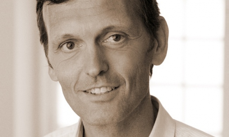
Eike Nagel
The comparatively fast method makes it possible to examine patients at an early stage and may prevent cardiac insufficiency or even a heart attack. “Diseases such as HIV, kidney damage, rheumatic diseases or tumours often affect the heart either directly or as a side effect of therapy,” says Nagel, describing groups potentially at risk. The cardiologist is convinced: “Nowadays we can treat or even cure so many diseases, but the heart suffers too and this should be carefully monitored as it mostly remains undetected.”
MRI is a non-invasive and gentle examination technique, which is less risky but just as efficient as an examination using a conventional heart catheter, where a thin tube is pushed in the direction of the heart through an artery. Nagel’s research group was recently able to demonstrate this in a large international multi-centre study that was met with international acclaim.
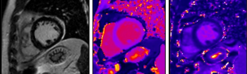
The Institute for Experimental and Translational Cardio Vascular Imaging also has state-of-the-art computer tomography equipment at its disposal that can produce three-dimensional images of the heart. These especially reveal calcium deposits and plaques in the artery walls which could rupture and trigger a sudden heart attack. “This allows us to determine the risk of a heart attack and the need for therapy fast and at an early stage, which can then be non-invasive,” says Nagel. Which technique is best for which patient is one of the research topics Nagel’s group is evaluating. In some patients, both may be needed and the Institute is optimally equipped to answer most aspects of heart disease thanks to its deep insight into the heart.
Nagel finds these rapid advances in imaging over the last decades fascinating: “Nowadays we can spot the slightest changes and literally get a clear picture of the heart’s condition.”
Source: Goethe-Universität Frankfurt am Main
10.12.2017