© Horacio Selva – stock.adobe.com
News • Assessment of spatial configuration
AI scans pathology slides to predict pancreatic cancer recurrence
Research insights could be used to guide more individualized surveillance, adjuvant therapy and clinical trial design
Mayo Clinic researchers have found that artificial intelligence (AI)-enabled spatial analysis can identify patterns in routine pathology slides that may help clinicians identify which patients with pancreatic cancer are at greater risk of recurrence after treatment and surgery.
The study, published in Clinical Cancer Research, suggests that looking at how residual cancer is organized — not just how much remains — could eventually improve estimates of recurrence risk, particularly for patients whose tumors show only a limited response to chemotherapy before surgery.
Current pathology assessments largely tell us how much tumor is left after treatment. We wanted to know whether the geography of that remaining cancer could reveal additional biology about recurrence risk
Ryan Carr
Patients with a more fragmented, intermixed pattern of cancer and the surrounding tissue, or stroma, had earlier recurrence. The amount of residual cancer alone did not reliably separate patients at higher and lower risk.
"Current pathology assessments largely tell us how much tumor is left after treatment. We wanted to know whether the geography of that remaining cancer could reveal additional biology about recurrence risk," says Ryan Carr, M.D., Ph.D., a Mayo Clinic oncologist and senior author of the study.
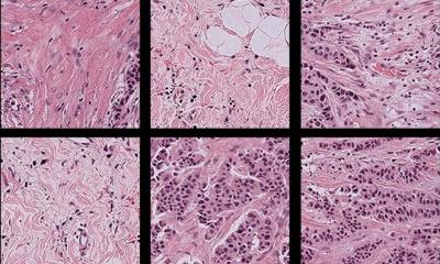
Dr. Carr and his team used an AI tool to identify cancer and stromal regions on standard pathology slides, then measured how those regions formed patches, boundaries and mixed areas to evaluate spatial patterns tied to a higher risk of recurrence.
The study analyzed tissue from 203 patients with pancreatic ductal adenocarcinoma who received treatment before surgery but showed only a limited pathologic response. The researchers combined an AI-enabled digital pathology platform with methods adapted from landscape ecology to analyze standard hematoxylin and eosin, or H&E, slides. The approach measured tissue shape, fragmentation, and the degree to which the cancer and stroma were intermixed.
The work builds on Dr. Carr's broader research applying ecological principles to cancer. His team uses machine learning and spatial analysis to map the pancreatic cancer ecosystem and study how cancer cells interact with neighboring cells and tissues — relationships that may influence treatment resistance and recurrence.
Two spatial signatures predicted disease-free survival even after accounting for stage, lymph node status, and other established clinical and pathologic risk factors. In one model, high-risk patients had a 71% higher adjusted risk of recurrence. In another, high-risk patients had more than twice the adjusted risk. These spatial models helped distinguish patients at higher and lower risk when standard measures, including how much cancer remained, did not.
Because the approach uses pathology slides already generated as part of routine care, it could potentially give clinicians additional information to inform recurrence risk without requiring another tissue test.
"What is exciting is that this information is already present in the tissue," Dr. Carr says. "AI-enabled analysis gives us a way to measure features that are difficult to capture by eye and potentially add another layer of precision to how we assess risk after surgery."
The research also found that high-risk spatial patterns contained fewer immune cells within the cancer itself, with immune cells tending to collect around the tumor instead of entering it. The finding underscores the importance of the tumor microenvironment — the cells and tissues surrounding a tumor — in treatment resistance and disease behavior.
More broadly, the work aligns with Mayo Clinic's Precure Research priority to use data and technology to predict risk earlier and create opportunities to intercept serious disease before it advances. "Our long-term goal is to better identify which patients remain at greatest risk and ultimately use that knowledge to guide more individualized surveillance, adjuvant therapy and clinical trial design," Dr. Carr says.
The researchers say the results are promising but need to be confirmed in prospective studies before this approach could be used to inform clinical decision-making.
The research was supported in part by the Gerstner Family Foundation Career Development Award, the Grand Forks Career Development Award, the Mayo Clinic Center for Clinical and Translational Science, and the ARPA-H ADAPT program. For a complete list of authors, disclosures and funding, review the study.
Source: Mayo Clinic
21.09.2026