Image source: University of Jyväskylä; photo: Petteri Kivimäki
News • dMMR prediction
AI helps analyse colorectal cancer samples
Researchers at the Faculty of Information Technology at the University of Jyväskylä have used artificial intelligence to speed up the analysis of colorectal cancer samples and predict the functioning of the cells’ DNA repair mechanism.
The AI model's analysis can help shorten diagnosis times, reduce costs, and improve the analysis's accuracy. The research, published in the journal Computer Methods and Programs in Biomedicine, was conducted in collaboration with the Central Finland Welfare Region and is co-funded by the European Union.
Analysing a cancer sample in a pathology laboratory–regarding, for example, the MMR mechanism–can take several days, whereas artificial intelligence can reduce the analysis time to minutes
Liisa Petäinen
The cell’s own error-correction mechanism, the so-called MMR mechanism, corrects small errors that occur during DNA replication. If this mechanism does not function properly, it can affect both cancer development and treatment decisions.
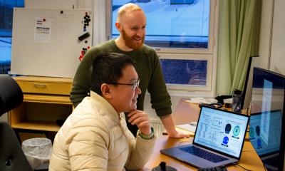
Liisa Petäinen, who led the study at the University of Jyväskylä, explains that analysing tissue samples is routine but time-consuming work. “Analysing a cancer sample in a pathology laboratory–regarding, for example, the MMR mechanism–can take several days,” says Petäinen, “whereas artificial intelligence can reduce the analysis time to minutes.”
Faster analysis could lead to cost savings and shorten the time it takes for a patient to receive a diagnosis and access treatment. At the same time, it would free up pathologists’ time for other tasks.
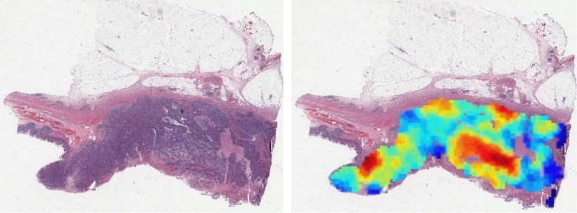
Image source: University of Jyväskylä
The analysis of cancer samples is currently based on assessments by pathologists, which makes the process highly manual and time-consuming. According to Petäinen, this is precisely where artificial intelligence can help. Typically, the analysis is conducted using twentyfold magnification of the tissue sample, but the researchers also tested AI-assisted analysis using a considerably broader fivefold magnification.
The researchers have reported that the model also performed reasonably well at this scale. Petäinen is hopeful that, in the future, tissue samples could be analysed in a single step using artificial intelligence. Analysing the entire tissue sample instead of only the tumour area would speed up screening, as the tumour area would no longer need to be identified separately in the image beforehand. The study also suggests that tissue features surrounding the tumour may help predict the function of the repair mechanism. Analysing the entire sample could therefore further improve the accuracy of the analysis.
The study was conducted together with pathologists and colorectal cancer experts from the Central Finland Biobank and the Wellbeing Services County of Central Finland. The dataset consisted of approximately 1,300 colorectal cancer patients from Central Finland. The model was also tested using data from Oulu University Hospital and the United States.
Tiina Jokela explains that Finland has high-quality biobanks, registers, and a unified healthcare system, which enable high-level research and faster implementation of results. “Central Finland offers a good pilot environment where research and clinical work can collaborate flexibly,” says Jokela. “The Central Hospital Nova of Central Finland provides clinical data and practical requirements for the research, while JYU provides its expertise in artificial intelligence and data analytics.”
According to the researchers, new methods need to be validated using larger datasets, as was also done in this study.
The study is part of Central Finland’s project AI-Hub II, which is co-funded by the European Union, and it was conducted in collaboration with the University of Jyväskylä, the Central Finland Biobank, and the Wellbeing Services County of Central Finland. The study was also funded by the Regional Council of Central Finland.
The model was trained using data from the project Colorectal Cancer in Central Finland 2000–2015. The development of the model has also utilised a cancer detection model developed in the Central Finland projects AI Hub I and II.
Source: University of Jyväskylä
29.05.2026